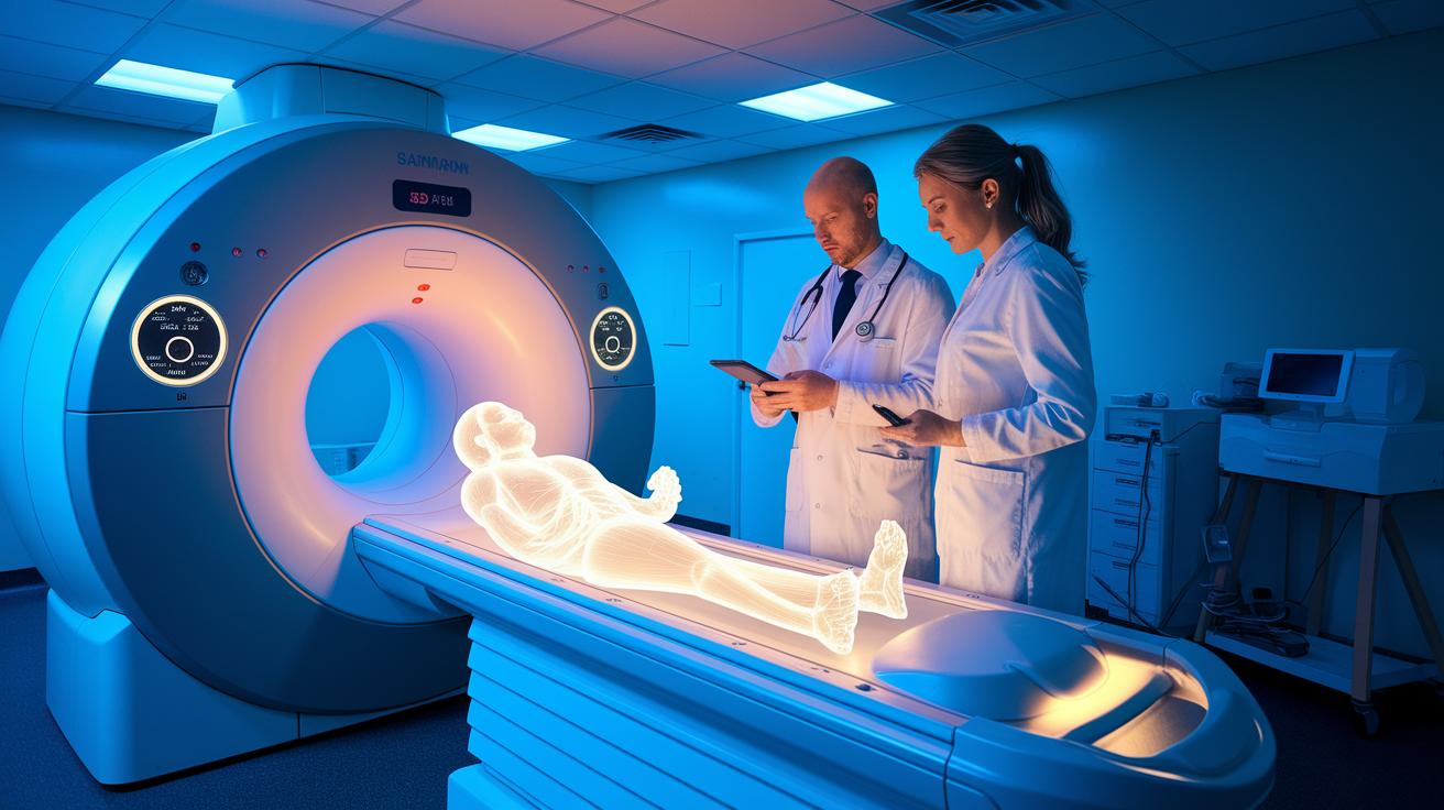

Could AI help spot issues in medical scans before a doctor does? New computer tools learn to read X-rays, CT scans, and MRIs like experienced professionals. They pick up on small details that might be missed by the human eye, which can lead to faster and safer diagnoses.
This technology changes raw data into clear insights, helping imaging tests become more efficient and easier to access. The improvements could mean better care for patients and a shift in today’s healthcare approach.
Read on to learn how AI is quietly transforming the world of medical diagnostics for the better.
AI in medical imaging explained: Advancing Diagnostics
AI in medical imaging uses computer programs that learn from data to study scans like X-rays, CTs, MRIs, ultrasound, and multispectral images. These tools spot patterns in pictures that the human eye might miss. For example, an AI-processed CT scan can show details that traditional methods might not catch, much like finding hidden art in an old building.
By analyzing huge amounts of data, AI can deliver fast and consistent results. Algorithms trained on large datasets pick up even tiny changes in a scan every single time. This means that AI can automatically point out tumors and other irregularities much quicker than a human expert.
AI also helps to cut down the time needed for scans and reduces patients’ exposure to ionizing radiation. Shorter scans and less radiation make the process safer and more comfortable. Plus, AI makes imaging easier to access in places where there aren’t many specialists, as it can help with screening and report creation.
Researchers are constantly updating these systems to keep them in line with the latest clinical standards. This ongoing work lays the groundwork for deeper use of AI in diagnosing illnesses, which can improve workflows and lead to better outcomes for patients.
Unpacking AI Techniques in Radiology Imaging
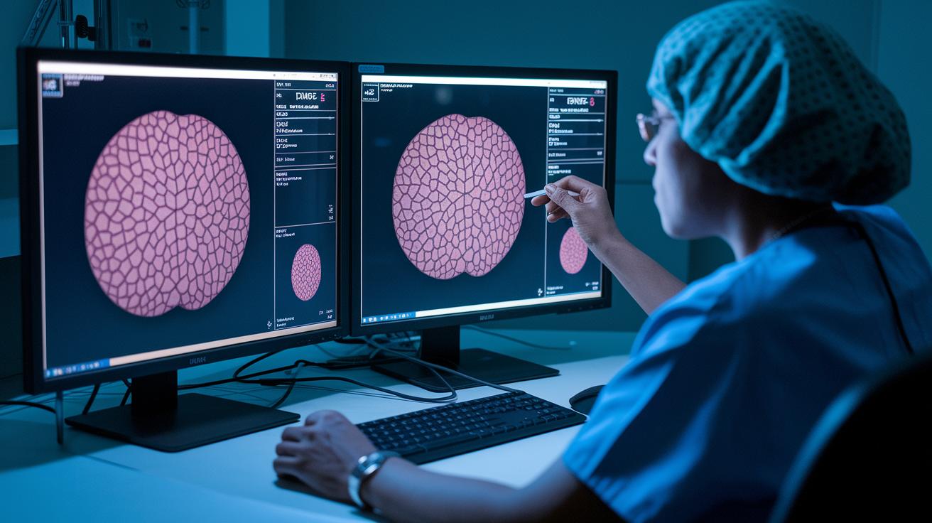
AI is changing how we look at medical images. Tools like convolutional neural networks (CNNs), random forests, support vector machines, and generative algorithms help break down complex scans into clear details. These models handle tasks such as dividing images into segments, rebuilding blurry scans, and cutting out unwanted noise. For instance, the U-Net model can quickly outline a tumor in a scan, much like a chef chopping vegetables. This quick approach is very helpful in cancer care, where fast measurements of tumors are essential.
These AI tools take the raw data from scans and turn it into clear, useful information. One important method, called radiomics feature extraction, picks out tiny details in images that are hard to see with the naked eye. This technique is valuable not only for cancer studies but also for heart, metabolism, and bone-muscle conditions. It helps doctors spot small textures and patterns that can guide decisions on treating diseases or planning care.
When these methods are combined in algorithmic image processing, they boost image quality and help doctors make better diagnoses. By reducing noise and bringing out key features, the images become clearer and more consistent. Using these AI approaches in radiology leads to faster reviews and deeper insights into a patient’s condition, setting the stage for care that is more personal and effective.
These advanced tools are transforming radiology, letting us measure images in ways that were once out of reach.
Integration of AI into Radiological Workflows
AI is reshaping everyday work in radiology. It automates common tasks such as marking images, sorting cases by urgency, writing up reports, and pulling information from patient records. This shift lets radiologists spend more time on the parts of care that truly matter.
Imagine a busy radiology ward where a computer quickly reviews images, highlights urgent cases, and prepares detailed reports. With these tasks handled automatically, staff can focus on direct patient care.
AI also plays a key role in screening. It helps check if patients meet the criteria for tests, looks for signs of disease, and tracks how well treatments are working. At busy or understaffed centers, this means every patient gets the attention they need.
The benefits are clear:
| Benefit | Description |
|---|---|
| Automated tasks | Repetitive work like image annotation is handled by AI. |
| Better screening | Improved checks on patient eligibility and disease detection. |
| Reliable data | Consistent imaging results improve trial quality. |
By streamlining these processes, radiology teams can start studies sooner and make decisions faster. This not only boosts overall efficiency but also helps speed up clinical and trial decisions that affect patient outcomes.
AI-Driven Applications Across Imaging Modalities
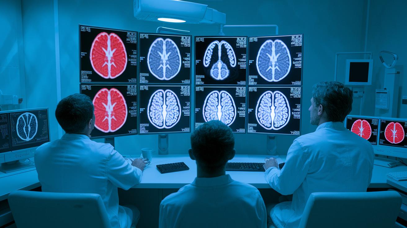
New AI tools are changing how CT and MRI scans help doctors make better decisions. These systems can spot coronary artery calcification and early signs of neurodegeneration that traditional methods might miss, giving doctors a chance to treat problems sooner.
In heart imaging, AI plays a key role. It automatically reviews echocardiograms, CT angiography, and cardiac MRI scans to show where the heart has structural issues or reduced blood flow. When a scan hints at a problem, like a slight narrowing of a blood vessel, it alerts doctors quickly so that urgent care can be provided.
Ultrasound imaging is also getting a boost from AI. Automated image reviews speed up exams and improve image clarity, which means that busy clinics can get consistent, reliable results every time.
X-ray work is similarly enhanced by AI. Tools now examine X-rays to highlight important features, reducing the risk of missing details. This helps radiologists focus on more complex cases while ensuring that nothing important is overlooked.
The DeepView AI System offers a detailed, pixel-level look at burn wounds. Multiple studies have shown that it accurately tracks healing progress, measuring small changes in tissue over time. This precision supports quicker, more effective treatment decisions.
Real-World Case Studies in AI for Medical Imaging
Aidoc's AI tool helps emergency teams act quickly by flagging critical conditions like a collapsed lung. In busy hospitals, the system sends real-time alerts that guide doctors to focus on cases that need urgent care. For example, it can detect hidden signs of a collapsed lung in time to save lives.
DeepView AI has developed a burn imaging platform that measures injuries at the pixel level. Backed by clinical studies from ABA, EBA, and BBA, this tool provides clear data on burn severity. It moves beyond visual estimates and offers reliable measurements, helping care teams adjust treatment plans effectively.
AI is also improving oncology research through automated segmentation. This process swiftly outlines tumors and lesions, cutting down hours of manual work to just minutes. The precision of AI segmentation helps study sponsors make timely, data-driven decisions during crucial stages of trials.
Overall, these case studies show that integrating AI into medical imaging can speed up diagnosis and treatment while keeping processes consistent. This leads to smoother hospital workflows and ultimately better outcomes for patients.
Benefits and Performance Metrics of AI Imaging
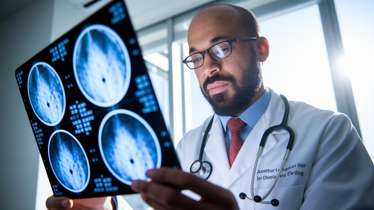
AI in medical imaging helps doctors see small changes in scans by highlighting details that are easy to miss. It picks up on minute pixel patterns and slight shades in imaging, which can make a big difference in spotting a disease early. One study showed that using AI cut the review time from 10 minutes to 5 minutes, which clearly demonstrates its usefulness in saving time.
AI also makes the reading process more consistent. By following a set evaluation process, it minimizes differences between different readers. This means patients can expect reliable and timely diagnoses. Clinical evaluations have noted that this uniformity leads to shorter reading times and fewer mistakes, which is a win for both care and efficiency.
Overall, these improvements lead to important gains in performance. AI-enhanced imaging workflows boost processing speed and result consistency, helping to improve patient care while keeping costs in check.
| Performance Metric | Impact |
|---|---|
| Sensitivity & Specificity | Better at picking up small details in images |
| Processing Time | Faster scans due to automated analysis |
| Error Rates | More consistent readings among different reviewers |
Challenges, Ethics, and Regulatory Considerations in AI Imaging
AI systems in medical imaging show promise but also have hurdles to overcome. Some of these tools are already approved and validated, while many are still in development or waiting for full regulatory clearance. This means it is not always clear which AI systems can be trusted for critical diagnostic tasks.
Getting AI tools to work in everyday clinical settings is not simple. Manufacturers must partner closely with healthcare providers to make sure these systems integrate well and do not slow down or disrupt workflows. For instance, a hospital might face problems shifting from manual to AI-based image annotations if the new system does not blend easily with the software already in use.
There is also concern about bias in these algorithms. Without training on diverse data and careful testing, AI tools may favor one group over another, resulting in unequal care. It is equally important to follow data privacy rules to protect patient information during analysis, which helps build trust in these emerging technologies.
- Some AI systems are approved while others await full regulatory clearance.
- Integrating AI tools with current clinical workflows can be challenging.
- Bias risks highlight the need for diverse data and thorough testing.
- Upholding data privacy is essential to protect patient information.
Future Directions and Emerging Trends in Medical Imaging AI
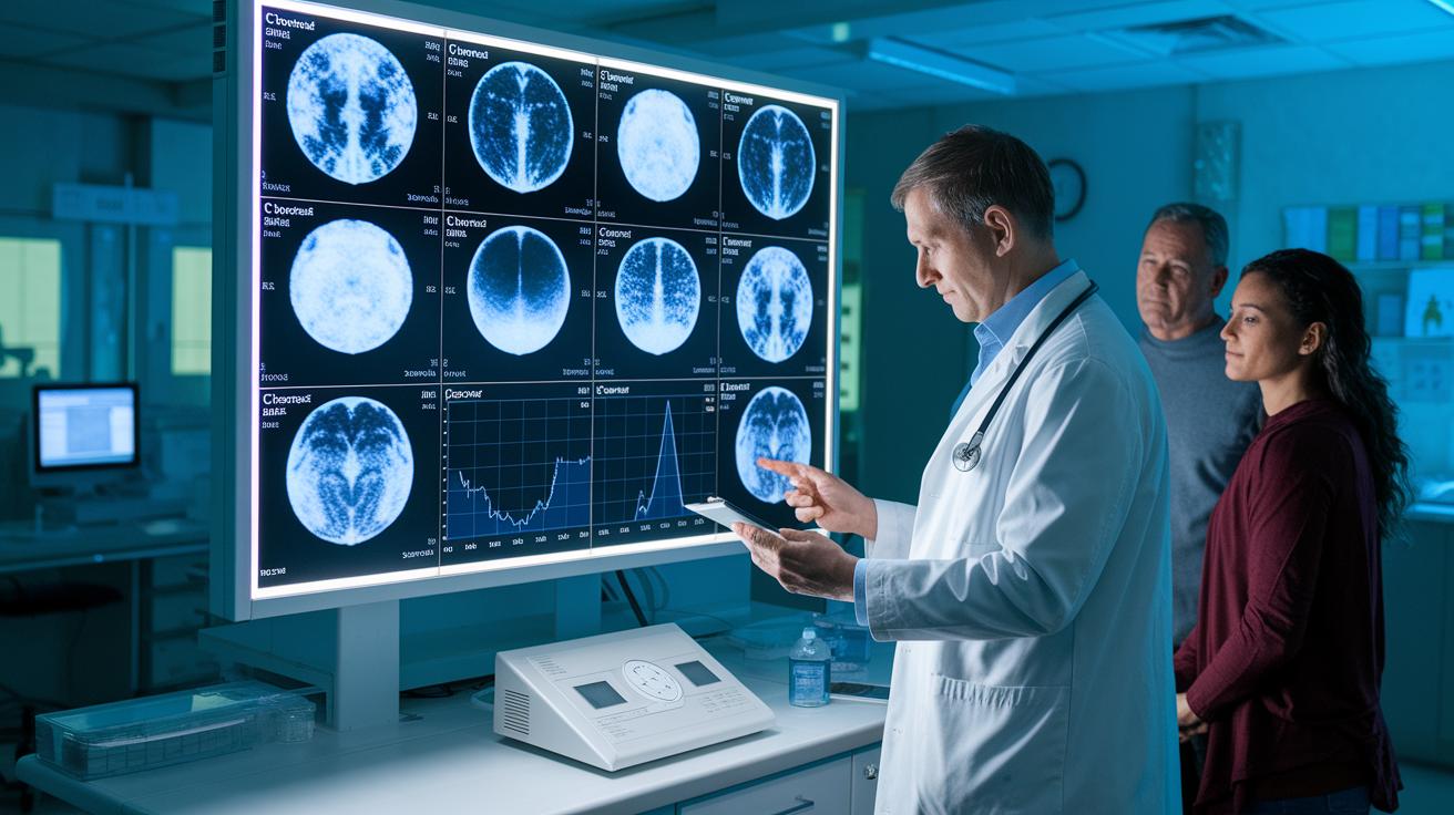
Recent studies show that imaging technology is moving fast and changing the way AI helps in medicine. Researchers are trying new methods that let computers spot patterns without needing lots of labeled data. For example, one study found that better models can learn from small sets of images, much like a student grasps a new idea after just a few examples.
Another promising trend is merging different types of images. By combining CT scans, MRIs, and digital pathology slides, doctors can get a fuller picture that helps them make more accurate diagnoses. At the same time, digital pathology analytics is developing tools that predict patient outcomes and offer more personalized care, paving the way for earlier interventions.
New techniques in quantitative analytics and image reconstruction are set to bring even clearer details about tissues and diseases. This means clinicians will have solid data to create treatment plans that fit each patient better. Real-time diagnostic systems and predictive models are also on the rise, allowing healthcare teams to make quicker decisions and improve patient care.
Final Words
In the action, the article broke down how AI in medical imaging explained key concepts and techniques. It described AI methods, real-world uses, and how these advanced tools fit into daily clinical routines. It also touched on benefits, performance metrics, and challenges such as ethics and regulation.
The piece offers a clear snapshot of emerging trends that promise faster, more consistent diagnostics. The journey ahead remains promising, providing a pathway to smarter, more personalized healthcare decisions.