
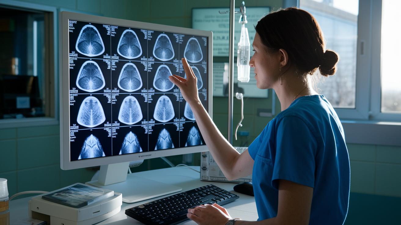

A new approach in computer vision is helping doctors catch tiny lesions before they become a bigger problem. Hospitals start by cleaning up digital images to remove any background noise and boost the contrast so that even the smallest details stand out. Then, smart computer systems work to spot and follow changes that could indicate disease. This technology makes medical images clearer and helps doctors decide on care faster and with more confidence.
Comprehensive Overview of Computer Vision in Radiology
Computer vision in radiology is changing how we work with medical images. First, hospitals load digital imaging files (called DICOMs) into the system. Next, these images are prepared in a step that fixes their orientation, cuts down background noise, and boosts contrast so details become clearer. For example, increasing the contrast in one x-ray can uncover small lesions that might go unnoticed.
After this cleaning step, the images are analyzed by computer algorithms. These systems use methods like convolutional neural networks for classifying images and U-Net type layouts for splitting images into important parts. This process helps the system automatically spot lesions and recognize key patterns in the images.
During this analysis, the system also maps how a disease changes over time in a patient. Special tools, like GANs and VAEs, are used to create realistic images when there isn’t enough real data. These computer-generated images help represent groups and conditions that are seen less often, and they also make it safer to share data while following privacy rules like HIPAA and GDPR.
Finally, a post-processing step smooths the image edges and removes unwanted marks. Together, these steps form a smooth workflow that speeds up exam reviews and improves diagnostic accuracy. By linking data loading, careful image preparation, smart analysis, and fine-tuned finishing touches, computer vision makes radiology faster, smarter, and more reliable.
Pipeline of Computer Vision in Radiology: From Data Ingestion to Clinical Integration

The process starts by gathering imaging files in DICOM format from PACS and converting them into a user-friendly format. This is like collecting all your ingredients before you start cooking.
Next, the images are preprocessed. Their spatial orientation is adjusted and different views are aligned. Techniques to reduce noise and enhance contrast help bring out important details. Even a small error here can have a big impact later. For example, a tiny misalignment might make an algorithm treat a small lesion like background noise, much like reading a map that's slightly rotated.
Then, spatial standardization corrects any drift or image issues, such as an upside-down brain scan that might confuse the detection model. This step makes sure images from different scanners match up properly before moving on to detailed analysis.
After standardizing the images, detection models take over. Some use one-stage networks similar to YOLO-style methods, others rely on two-stage region proposals, or start with segmentation. Each technique has its own strengths in detecting anatomical details and anomalies in radiographs.
Next, post-processing fine-tunes the results through methods like thresholding and morphological adjustments. These tweaks need to be handled carefully because even a small error, like an inaccurate segmentation, could exaggerate the size of a tumor.
Finally, the system integrates with electronic health records and includes a review by radiologists. This stage is still evolving, but adding clinical context is key to ensuring that image analysis remains accurate and reliable.
Deep Learning Models and Imaging Analysis Algorithms in Radiology
Deep learning models power today's radiology computer vision tools. These systems help find important details in images. For example, convolutional neural networks (CNNs) learn from pixel patterns to tell apart healthy lung tissue from early signs of infection.
U-Net segmentation networks are built to mark tissue edges. They work very well with CT and MRI scans, making them useful for spotting lesions and outlining organs. This precision helps doctors monitor tumor size over time.
Generative models like GANs (Generative Adversarial Networks), VAEs (Variational Autoencoders), and diffusion models can produce high-quality synthetic images. These images help train AI systems, especially when real patient data is limited for rare diseases. One study found that synthetic images generated by GANs boosted model performance by mimicking rare and complex disease details. This process not only increases the amount of training data but also prepares the system for a range of clinical scenarios.
Transfer learning is another useful approach. It lets developers fine-tune models that were pre-trained on large datasets using smaller, specific groups of images. This cuts down the need for vast amounts of labeled medical images and speeds up the creation of diagnostic tools.
| Model Type | Primary Use | Key Advantage |
|---|---|---|
| CNN | Image classification | Learns features in a hierarchy |
| U-Net | Semantic segmentation | Delivers precise boundaries |
| GAN/VAE | Synthetic data generation | Increases data variety |
Together, these techniques improve diagnostic accuracy in radiology by addressing challenges in image classification, segmentation, and data expansion.
Clinical Applications of Computer Vision in Radiology
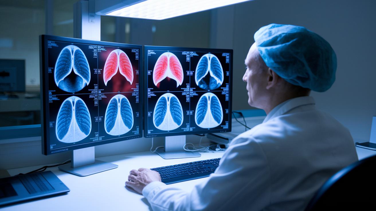
Computer vision is changing how radiologists spot and treat health issues. New systems can now find lung nodules on chest CT scans with 15% better sensitivity. One study found that these algorithms pick up tiny nodules that might have been missed before, making early treatment a real possibility.
Digital x-ray algorithms use advanced contrast techniques to keep important diagnostic details visible while lowering radiation exposure. In practice, when a technician adjusts an image, the software fine-tunes the details and highlights subtle issues, without extra risk to the patient.
CT scan tools now use 3D region-proposal networks to help detect liver lesions and monitor tumor size changes over time. These systems provide clear, measurable data that shows whether a tumor is growing or shrinking, which helps doctors decide on the best treatment.
Modern MRI classification models can distinguish between different tissue types with improved accuracy. They aid in evaluating brain tumors and spotting signs of multiple sclerosis. This detailed information allows doctors to create treatment plans that fit each patient’s needs.
Predictive analytics add another layer to diagnosis by tracking changes in radiographic biomarkers over time. These tools sort patients by risk based on how these markers evolve, so clinicians can focus on those in need of immediate attention.
The key benefits include faster exam reviews and more accurate diagnoses. By integrating these advanced algorithms into daily radiology work, doctors can better interpret images and provide focused care. This progress is paving the way for a future where data plays a central role in diagnostic imaging.
Challenges in Radiology Computer Vision: Bias, Interpretability, and Error Patterns
Radiology computer vision systems can make mistakes that affect how accurately they find problems in scans. One common issue is that these systems sometimes miss small details because they get confused by overlapping shadows or medical device artifacts. For example, an algorithm might not spot a tiny lung nodule because the shadow from a device looks very similar to a real lesion. This kind of error can delay treatment and affect patient care.
Models are often trained on scans from a single institution. When they are used on images from other hospitals, differences in equipment and scanning protocols can create extra challenges. This situation, known as domain shift, leads to inconsistent results that confuse the detection process.
Another problem is protocol drift, where changes in scanning practices over time are not reflected in the training data. When the frequency of disease in training images does not match that in real life, the output may be off. To help overcome these challenges, experts are turning to explainable AI. Such systems show exactly which areas of an image were flagged as abnormal and offer a simple explanation for their decisions. While this clarity improves understanding for radiologists, it also adds extra work for the computer systems.
Overall, continuous testing on varied data and steady collaboration among experts are vital to making these tools more reliable and safe for everyday use.
Regulatory and Ethical Considerations for AI-based Radiology Tools
Privacy rules like HIPAA and GDPR mean that health images must have personal details removed or be replaced with synthetic versions before sharing. This step protects patients and builds well-organized image collections for clinical use. One radiology team expanded its training set using synthetic data, avoiding privacy issues while keeping the images realistic, similar to swapping a sensitive ingredient for a safer one in a recipe.
Different institutions often use varied data systems and file formats, which delays training AI models across centers and makes testing algorithms tougher. To overcome these obstacles, experts are working on methods to boost datasets and check their quality. These efforts help ensure that synthetic images closely mimic real ones, improving medical imaging results.
New rules now consider AI software as a medical device. This change means there must be a clear certification process with careful testing and detailed documentation. At the same time, strong data management is key to meeting these challenges. Overall, ethical oversight and solid quality measures are essential for safely using AI tools in clinical settings.
Future Directions: Predictive Healthcare Imaging with Computer Vision
Integrating imaging with patient records and gene profiles is changing how doctors diagnose illnesses. By combining X-rays, lab results, and health records, these systems create a complete picture of a patient’s condition. This mix of data helps spot even small changes before serious problems arise. For example, linking radiography with lab tests can reveal early signs of disease.
Real-time imaging is also moving forward. In operating rooms and emergency settings, fast image processing helps guide urgent clinical decisions. Cloud-based solutions and learning models that use data from multiple centers protect patient privacy while building smarter systems. This collaborative approach makes it possible to adjust quickly as new trends emerge.
Researchers are now developing systems that learn continuously while using safeguards to avoid mistakes over time. These advancements promise quicker, more precise diagnostic insights during critical moments. Ongoing studies and pilot projects are steadily refining these tools, aiming for even greater accuracy and faster turnaround in patient care.
Final Words
In the action from data ingestion to clinical integration, the blog post unpacked how computer vision in radiology boosts diagnostics from image classification to predictive analytics. It took us through key model stages, technical challenges, and regulatory concerns, offering clear insights on overcoming common pitfalls.
The discussion also highlighted emerging trends, including multimodal data fusion and real-time imaging. This exploration urges a positive outlook as innovative tools steadily improve healthcare imaging and support better-informed clinical decisions.
FAQ
What is computer vision in medical imaging?
Computer vision in medical imaging uses AI algorithms to analyze radiographs. It handles tasks like lesion detection and segmentation, helping speed up diagnosis and improve accuracy in interpreting images.
Why isn’t AI replacing radiologists?
AI isn’t replacing radiologists because it works as an aid rather than a standalone solution. Radiologists still provide essential judgment, integrate clinical context, and manage complex decision-making.
What are computer vision medical imaging projects?
Computer vision medical imaging projects develop AI pipelines to process imaging data. They focus on tasks like data ingestion, image preprocessing, and pattern recognition to support radiologists in their workflow.
What is computer vision in health care?
Computer vision in health care integrates AI-driven image analysis into clinical processes. It improves diagnostic speed and accuracy across different medical fields, including radiology and pathology.
How does deep learning-enabled medical computer vision work?
Deep learning-enabled medical computer vision employs techniques such as convolutional neural networks and U-Net segmentation. These methods boost the precision of detecting anatomical structures and identifying pathological features.
What are biomedical computer vision projects?
Biomedical computer vision projects apply AI to analyze medical images. They support tasks like tissue segmentation and disease mapping, enhancing the ability to track and assess health conditions.
Where can I find deep learning in computer vision PDF resources?
Deep learning in computer vision PDFs offer detailed studies on neural network architectures and performance benchmarks. These resources explain the methods used to enhance imaging analysis in radiological and biomedical applications.
What does a review of tracking and mapping in medical computer vision cover?
Reviews on tracking and mapping in medical computer vision cover methods for image registration, error reduction, and integration into clinical workflows. They provide insights into improving pipeline performance from data ingestion to clinical use.
What is the highest paying medical imaging job?
The highest paying roles in medical imaging are typically specialized positions, often in radiology informatics or advanced clinical radiology. These roles combine high technical expertise with clinical decision-making skills.
Is computer vision a dead field?
Computer vision remains an active and evolving field. Continuous improvements in AI techniques keep the field innovative, driving advancements across radiology, manufacturing, and broader health-care applications.