

When minutes count, your doctor needs to choose the best imaging test. In emergencies, a CT scan is a trusted option. It quickly produces clear images of your bones and spots urgent issues in just minutes.
An MRI, on the other hand, offers a detailed look at soft tissues like the brain and joints. It takes a bit longer, but it can show finer details that a CT scan might miss.
Understanding the strengths of each test helps your doctor decide which one is right for you, ensuring you get the proper care when it matters most.
Direct Comparison of CT Scan vs MRI for Diagnostic Clarity
CT scans work by using X-rays to create images in around 5 minutes. This quick process makes them a go-to choice for emergency cases like trauma, checking for pulmonary embolism (a blood clot in the lungs), and looking at bone issues. Because the images are produced fast, doctors can quickly spot urgent problems and decide on the next steps. Board-certified radiologists carefully check these scans to make sure they meet high standards for clear diagnosis.
MRI uses strong magnets (1.5 Tesla) and radio waves to produce detailed images, but it takes 20 to 45 minutes. This extra time is worth it when doctors need a closer look at brain disorders, nerve problems, or injuries to muscles and joints. Modern wide-bore MRI machines are designed to speed up the process and help patients feel more comfortable, especially those uneasy in tight spaces. Expert radiologists, trained specifically in MRI, review these scans to ensure accurate results.
Choosing between CT and MRI depends on how urgent the situation is, the patient’s history, and what needs to be checked. CT scans are usually preferred when time is critical and details about bones or acute bleeding are needed. On the other hand, MRI is better for clear images of soft tissues, like in brain or joint exams. Matching the strengths of each scan to the patient’s condition lets doctors pick the best approach for every case.
Imaging Techniques in CT vs MRI: X-Ray Versus Magnetic Resonance
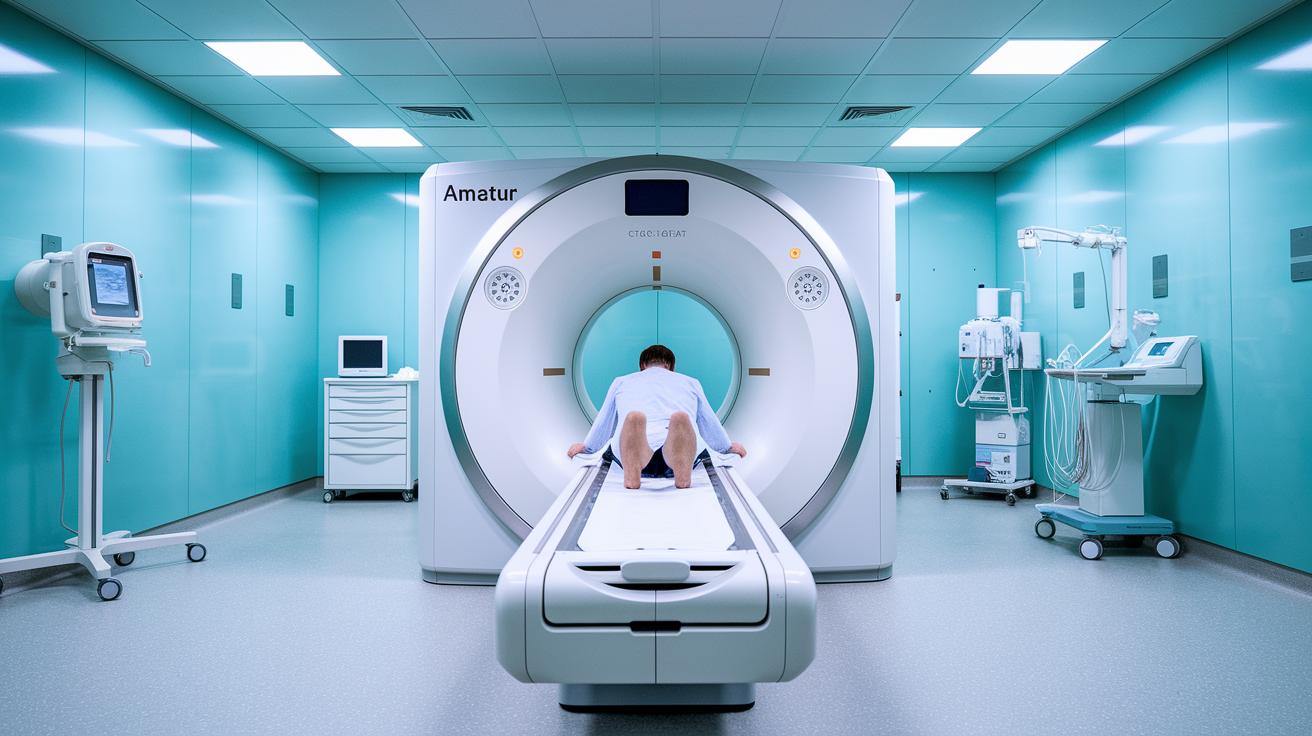
CT scanners work by rotating an X-ray tube and detectors to capture many thin slices of your body. These slices are then combined into a full 3D image. The exam usually gives off between 2 and 10 mSv of ionizing radiation and takes only a few minutes, making it a quick choice for spotting bone injuries, lung problems, or other urgent issues.
MRI machines, on the other hand, use a steady magnetic field (often 1.5 Tesla or more) along with bursts of radio waves to create images. This method measures how tissues relax (called T1 and T2 relaxation) and builds detailed, multiplanar 3D views of soft tissues like the brain and muscles, all without using ionizing radiation.
- CT uses radiation to create images, while MRI relies on magnets and radio waves.
- A CT scan is fast, usually around 5 minutes, compared to an MRI that can take 20 to 45 minutes.
- CT reconstructs multiple thin slices into a 3D picture, whereas MRI produces detailed 3D and multiplanar images of soft tissues.
CT Scan vs MRI: Preferred Clinical Applications by Body System
When doctors need quick answers in an emergency, they often choose CT scans. These scans help detect problems like acute strokes and head injuries because they produce fast images that guide urgent care.
For brain issues, MRI is usually the go-to test. Its clear images are great for spotting multiple sclerosis marks and spinal cord lesions, showing even the smallest details.
When it comes to bone problems, CT scans are very useful. They clearly show fractures and fine details in bones, which is important after head injuries or other skeletal issues.
For blood vessel concerns, CT angiography is a fast option to check for blockages. Meanwhile, MRI angiography adds extra information by showing how blood flows through the vessels.
In cases of joint or soft tissue injuries, MRI is preferred over CT. It gives a detailed look at cartilage, ligaments, and menisci, making it ideal for sports injuries and joint conditions.
For assessing heart health, CT scans are often used to measure coronary calcium and estimate heart disease risk. However, when doctors need to check heart muscle and overall tissue health, MRI is the better choice.
When evaluating tumors and organ problems, each scan has its strengths. CT scans quickly capture bone details and calcifications, which is helpful in emergencies when fast decisions must be made. MRI, with its detailed soft tissue images, offers clear pictures of tumors in organs like the breast, liver, or brain. This helps doctors understand how far a tumor has spread into nearby tissues, a key factor in planning treatment. By combining both methods, doctors can use the speed of CT with the detailed images of MRI to create a complete picture and design a treatment plan that fits the patient's needs.
Safety, Radiation Exposure, and Patient Comfort in CT vs MRI
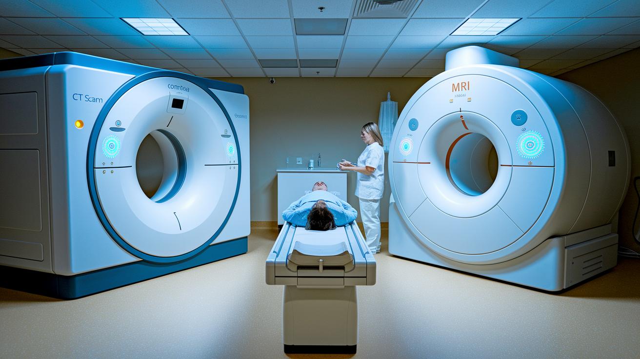
CT scans use a small amount of ionizing radiation, usually between 2 and 10 mSv per exam. Even though the overall risk is low when the scan is needed, radiation remains a consideration. MRI, on the other hand, uses strong magnetic fields and radio waves, so it doesn’t expose you to radiation. Each exam uses a special contrast agent to help produce clearer images. For CT scans, this is an iodinated contrast that comes with a 0.2% to 0.7% chance of causing an allergic reaction and may affect kidney function. MRI uses a gadolinium-based contrast with severe reactions reported in less than 0.04% of cases. However, people with kidney problems should be cautious because this contrast can, in rare cases, lead to a condition called nephrogenic systemic fibrosis.
| Modality | Contrast Agent & Risk |
|---|---|
| CT | Iodinated contrast; 0.2–0.7% allergic reaction, potential kidney issues |
| MRI | Gadolinium contrast; <0.04% severe reaction, caution with renal impairment |
Before the exam, talk with your doctor about any past allergies or kidney issues. For CT scans, your doctor may run blood tests to check your kidney function. If you’re scheduled for an MRI, let the team know if you’re uncomfortable in small spaces or sensitive to loud noises. Many facilities offer wide-bore machines, ear protection, or even music to help you relax. Following all pre-scan directions, such as removing metal items, will help ensure a safe exam and the best possible images.
Practical Considerations: Cost, Availability, and Workflow for CT vs MRI
- Cost: CT scans usually cost between $300 and $1,500, while MRI tests tend to range from $600 to $3,000.
- Scheduling: CT appointments are often available the same day and take about 15 minutes. In contrast, MRI slots might require a 1- to 2-week wait, and the tests last between 30 and 60 minutes.
- Throughput: A CT scanner can typically serve 20 to 30 patients each day, whereas an MRI machine usually handles 8 to 12.
- Insurance: Both tests are generally covered when they are medically necessary, although the preauthorization process may differ.
- Location: Many imaging centers offer both CT and MRI services in urban and suburban areas.
When choosing which test to schedule, consider more than just the cost. In emergencies, CT scans are often the best choice because they are available quickly and return results fast. Although MRIs provide more detail for soft tissues, they may require more planning due to longer exam times and fewer available slots. The daily patient flow and the clinic's workflow can also affect which test is best for your situation. It’s a good idea to talk with your healthcare provider about your specific needs, balancing urgency, cost, scheduling, and insurance details to get the most effective diagnosis.
Emerging Innovations and AI Integration in CT vs MRI Diagnostics
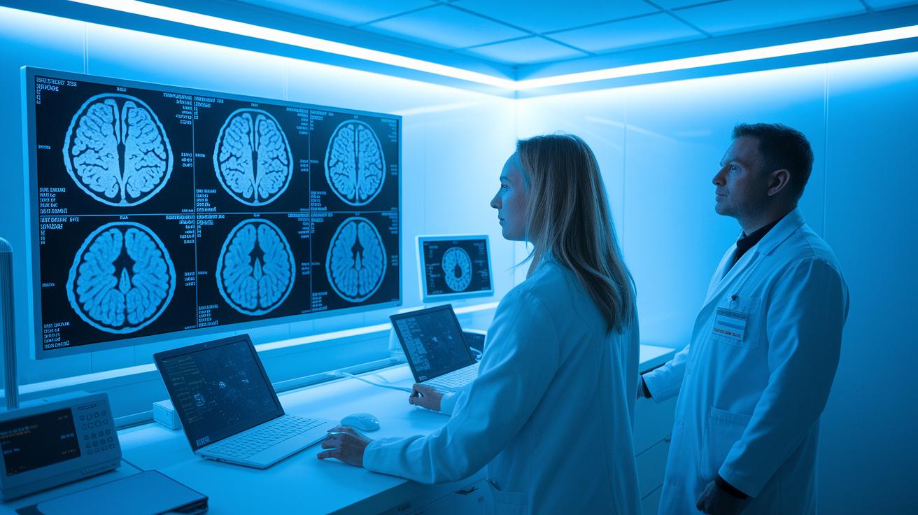
New advances in CT and MRI are speeding up scans and offering clearer images. Updated AI can now spot serious brain bleeds on CT scans within seconds, which helps doctors act quickly in emergencies. Deep-learning techniques have lowered the radiation dose by 30-50%, making scans safer without losing detail.
On the MRI side, methods like MR fingerprinting and compressed sensing have reduced scan times by up to 40%. This means patients spend less time in the scanner while still getting high-quality images of soft tissues. These improvements make patients more comfortable and boost the accuracy of diagnosing complex conditions.
AI is playing a big role in these upgrades:
- It detects brain bleeds quickly, improving emergency response.
- Deep-learning methods help lower CT radiation levels.
- MR fingerprinting delivers fast, sharp images of soft tissues.
- Compressed sensing cuts down MRI exam times significantly.
- Advanced tools now offer real-time 3D views and detailed tissue maps.
Final Words
In the action, our article compared CT scan vs MRI: which test is better for what by looking at speed, image quality, safety, and patient comfort. We broke down each method’s strengths in emergency settings, soft tissue detail, and clinical workflow. We also highlighted cost differences and emerging AI innovations that may shape future diagnostics.
Choosing the right test is about matching clinical need to each modality's unique benefits. Move forward with confidence, knowing that both scans offer valuable insights for better health decisions.
FAQ
What unique information do CT scans and MRIs provide that the other cannot?
CT scans reveal quick bone detail and acute trauma signs using X-rays, while MRIs offer high-resolution soft tissue images to evaluate the brain and nerves, giving complementary diagnostic insights.
How does MRI compare to CT for cancer detection?
MRI provides detailed soft tissue contrast crucial for tumor characterization, while CT scans offer rapid imaging of calcified masses and spread. Each modality is chosen based on the specific cancer type and clinical need.
Which imaging method is better for evaluating the brain?
MRI delivers superior soft tissue detail to detect subtle brain lesions and nerve changes, whereas CT scans are valuable for identifying acute bleeding and skull fractures in emergency settings.
Which imaging modality is considered safer, CT or MRI?
MRI is regarded as safer because it uses magnetic fields and avoids ionizing radiation, while CT scans expose patients to low levels of radiation, which are acceptable in urgent clinical situations.
How effective are MRI and CT scans in detecting nerve damage?
MRI is more effective in detecting nerve damage due to its detailed soft tissue contrast, whereas CT scans provide less clarity on nerves but remain useful when assessing surrounding bone injuries.
Which imaging method is preferred for evaluating the liver?
MRI is preferred for liver imaging because it offers better soft tissue resolution to identify lesions and tissue changes, while CT scans quickly assess liver structure during urgent evaluations.
Which modality is more effective for spine evaluation, MRI or CT scan?
MRI is favored for spine assessments because it clearly shows nerve compression and disc abnormalities, whereas CT scans are faster and better at depicting bony structures of the spine.
How can one differentiate between MRI and CT scan images?
MRI images feature smooth soft tissue contrast and less bone detail, while CT images provide sharper, well-defined outlines of bones and anatomy, aiding in distinguishing the two modalities.
Does MRI effectively show nerve damage?
MRI effectively displays nerve damage by highlighting soft tissue changes, inflammation, and lesions, making it a reliable tool for evaluating nerve injuries compared to CT scans.
Can CT scans reveal muscle inflammation?
CT scans can reveal muscle inflammation; however, their sensitivity is lower compared to MRI, which better differentiates soft tissue changes and subtle inflammatory signs.
Why do doctors sometimes prefer CT scans over MRIs?
Doctors often prefer CT scans for their rapid imaging, excellent bone detail, and reduced sensitivity to movement, making them ideal for emergency and trauma assessments.