


Worried that CT scans might be dangerous? The facts show they aren’t as risky as you might think. A head CT scan gives about 2 mSv of radiation, which is similar to what you receive from natural background radiation in less than one year. An abdomen or pelvis CT scan gives around 10 mSv, roughly the same as three years of everyday exposure. Doctors rely on CT scans because they provide detailed images that can quickly detect serious issues. Understanding these numbers can help ease concerns about the small amount of extra radiation involved.
CT scan radiation risk: how much exposure Reassures
CT scans use more ionizing radiation than regular X-rays or mammograms. A head CT gives about 2 mSv, while an abdomen or pelvis CT may expose you to roughly 10 mSv. For context, most people receive around 3 mSv per year from natural background radiation. This means a head CT is like getting less than a year’s natural exposure, and an abdomen CT can equal about three years of everyday background radiation.
A standard chest X-ray typically uses about 0.1 mSv, and a mammogram is under 0.5 mSv. While CT scans deliver more radiation, they are used because they provide detailed images that help doctors see inside the body clearly. This benefit usually outweighs the extra dose of radiation.
Radiation measurements are based on standardized imaging dosimetry, which helps patients and doctors discuss the risks and benefits with clear numbers. Keeping these figures in mind can lead to informed decisions about when such tests are necessary.
A quick recap:
| Type of Scan | Approximate Radiation (mSv) |
|---|---|
| Head CT | 2 |
| Abdomen/Pelvis CT | 10 |
| Chest X-ray | 0.1 |
| Mammogram | 0.5 or less |
| Annual Background Radiation | 3 |
Understanding these numbers helps you balance the small risk of extra radiation with the clear advantages of accurate medical imaging.
CT Scan Radiation Improvements and Comparisons
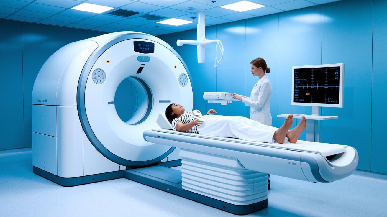
CT scans now use improved low-dose techniques that cut down on radiation while still delivering clear diagnostic images. Over the years, CT technology has grown safer, so today's exams use less radiation than older machines. For example, a head CT usually delivers about 2 mSv and an abdomen or pelvis CT around 10 mSv, and newer methods can reduce these levels even more.
Although CT scans give higher doses than chest X-rays or mammograms, they are still much lower than the exposure from nuclear events. These advances help doctors detect problems early while keeping patient safety in mind.
| Modality | Typical Effective Dose (mSv) | Equivalent Background Years |
|---|---|---|
| Chest X-ray | 0.1 | ~0.03 |
| Mammogram | 0.5 | ~0.17 |
| Head CT | 2 | ~0.67 |
| Abdomen/Pelvis CT | 10 | ~3.33 |
Understanding Health Risks of CT Scan Radiation
CT scans use radiation to create detailed images of the inside of your body. While this radiation might cause small changes in cells, the risk of developing cancer remains very low. We call this a stochastic risk because it is a matter of probability rather than certainty. Imagine each scan as tossing a coin, each has a slight chance of a specific outcome. One scan is like adding one drop of dye to a large bucket of water, almost unnoticeable on its own, but many drops can add up over time. For patients who need several scans, like during cancer treatment, the benefits of an accurate diagnosis and effective treatment planning outweigh the small, theoretical risk. Medical professionals also use lead shields and personal dosimeters to track and limit their own radiation exposure.
Stochastic Effects and Cancer Risk
Research shows that the chance of developing cancer from a single CT scan is very low. However, each additional scan adds a bit more radiation exposure, and that extra radiation can slightly increase the long-term risk. Doctors carefully weigh this small increase in risk against the clear benefits of accurate diagnosis. Each scan is reviewed to ensure that patients receive necessary information while keeping exposure as low as possible.
Deterministic Effects at Diagnostic Doses
In everyday CT imaging, the radiation dose is so low that it does not cause immediate tissue reactions like skin burns. There is a safe threshold below which these effects do not occur. Radiologists stick to strict guidelines and track radiation doses closely to keep exposure well below harmful levels. Before performing a scan, healthcare providers evaluate the benefits and any risks, ensuring that finding a health issue quickly is more important than the very minimal risk from the scan.
Identifying Patients Most Vulnerable to CT Scan Radiation

Some patients face extra risks from the small amounts of radiation in CT scans, especially when they need repeated exams. Women, young people, and those with chronic conditions can accumulate more radiation over time, which may raise their overall risk.
For instance, a young patient who must have many scans over several years will gather a higher dose of radiation. To reduce this risk, doctors often choose imaging methods that do not use ionizing radiation. Methods such as MRI and ultrasound are common choices because they use magnetic fields, radio waves, or sound waves instead of radiation.
Key groups at higher risk include:
- Women, who generally face a higher lifetime risk from radiation.
- Young individuals, whose growing tissues are more vulnerable.
- People with chronic conditions that require frequent scans.
- Individuals with a family history of radiation sensitivity.
To keep patients safe, doctors may suggest:
- MRI scans, which rely on magnetic fields and radio waves.
- Ultrasounds, which use sound waves for imaging.
These alternatives lower the overall radiation exposure while still giving the needed diagnostic insights. The focus is always on weighing the benefits of each test against the small, potential risks from repeated exposures.
Strategies and Protocols to Minimize CT Scan Radiation Dose
Modern CT scanners use built-in techniques to lower the radiation patients get while still producing clear images. New advances in detector technology and scan software have helped a lot with this progress. Radiology teams follow strict protocols and quality checks to keep patient radiation within safe limits. For example, many scanners automatically adjust the dose based on a patient’s body size, which makes each scan safer.
Here are five simple strategies used in many facilities:
-
Iterative reconstruction techniques: This method cuts down on image noise so that lower radiation doses can still produce quality images. A radiologist explained it like this: switching to this method is like swapping a foggy window for a clearer one, so less light (or radiation) is needed.
-
Automatic exposure control: These machines adjust the radiation dose based on the body part being scanned and the patient’s size, which makes sure the image is clear with the smallest necessary dose.
-
Low-dose scan modes: CT systems now offer settings that reduce radiation output, which is especially useful when patients need follow-up exams or repeated scans.
-
Quality assurance and dose tracking: Regular checks and dose tracking help make sure every exam meets safety standards. Technologists keep an eye on protocols to maintain the highest safety levels.
-
Advanced detector technology and optimized scan software: Improved hardware and smarter software mean that scans can be done with lower doses without losing important details, striking a better balance between diagnostic quality and patient safety.
Guidelines and Professional Recommendations on CT Radiation Doses

Leading organizations like the American College of Radiology (ACR) and the National Council on Radiation Protection (NCRP) set diagnostic reference levels (DRLs) for CT scans. These levels help ensure that radiologists use the smallest effective dose of radiation while still capturing clear images for accurate diagnoses.
Imaging centers keep a close eye on their CT doses by regularly reviewing their records. For example, a facility might check its dose logs every 3 months to make sure the scanner is within DRL limits. This ongoing review acts like a simple quality checklist to make sure scans stay safe.
Doctors also take time to explain these guidelines during patient consultations. They point out that the benefits of a CT scan far outweigh the small, controlled amount of radiation used. Patients learn that careful protocols are in place to reduce risks while helping doctors spot problems quickly.
These recommendations and regular checks give both patients and clinicians confidence in every CT exam. With consistent quality assurance and clear communication about dose limits, everyone can feel more secure knowing that the benefits of early diagnosis significantly outweigh the risks.
Final Words
In the action, the article explains CT scan radiation risk: how much exposure patients face during a CT scan.
It shows that CT doses, measured in mSv, far exceed those from X-rays while still falling under regulated limits.
The report reviews data that compares CT exposure with natural background and other imaging methods.
It also outlines risk factors and strategies to lower patient dose, supported by expert guidelines.
This clear summary empowers you to weigh risks and benefits, making informed decisions for better care.
FAQ
How to get rid of radiation after CT scan
The radiation from a CT scan typically clears naturally from your body over time, as the body eliminates ionizing particles. No special treatment is needed since the doses are managed and low for single scans.
How many CT scans are safe per year
The number of CT scans considered safe varies by individual and clinical need. For medically necessary exams, the benefit usually outweighs the small risks, though unnecessary repeated scans should be avoided.
CT scan radiation dose chart
The CT scan radiation dose chart shows that head CTs generally deliver around 2 mSv and abdominal scans up to 10 mSv, in contrast to a standard chest X-ray (~0.1 mSv) and natural background radiation (~3 mSv/year).
Minimum gap between two CT scans
The minimum gap between CT scans depends on the patient’s condition and clinical need. Radiologists often advise a gap of several months when possible to minimize cumulative radiation exposure.
CT scan cancer risk calculator
A CT scan cancer risk calculator estimates potential long-term cancer risk based on the radiation dose received and cumulative exposure, serving as a guide rather than a definitive prediction tool.
Can one time CT scan cause cancer
A one-time CT scan delivers a low level of radiation that poses a theoretical, minimal cancer risk, with diagnostic benefits outweighing the risk when the scan is medically indicated.
CT scan radiation side effects
CT scan radiation side effects mainly involve a small, theoretical increase in long-term cancer risk from cumulative exposure, with no immediate side effects like skin reactions at diagnostic dose levels.
Is one CT scan harmful
One CT scan is generally not harmful because the radiation dose is comparable to a few years of natural background exposure, and the immediate diagnostic benefit usually surpasses the minimal risk.
How much radiation does a CT scan expose you to
A CT scan exposes you to between 2 and 10 mSv of radiation depending on the area scanned, which is several times higher than standard X-rays but still within controlled and safe limits for clinical use.
Does a CT scan take years off your life
A single CT scan is unlikely to reduce lifespan. The small cumulative radiation risk assigns a theoretical probability of future cancer rather than an immediate impact on life expectancy.