
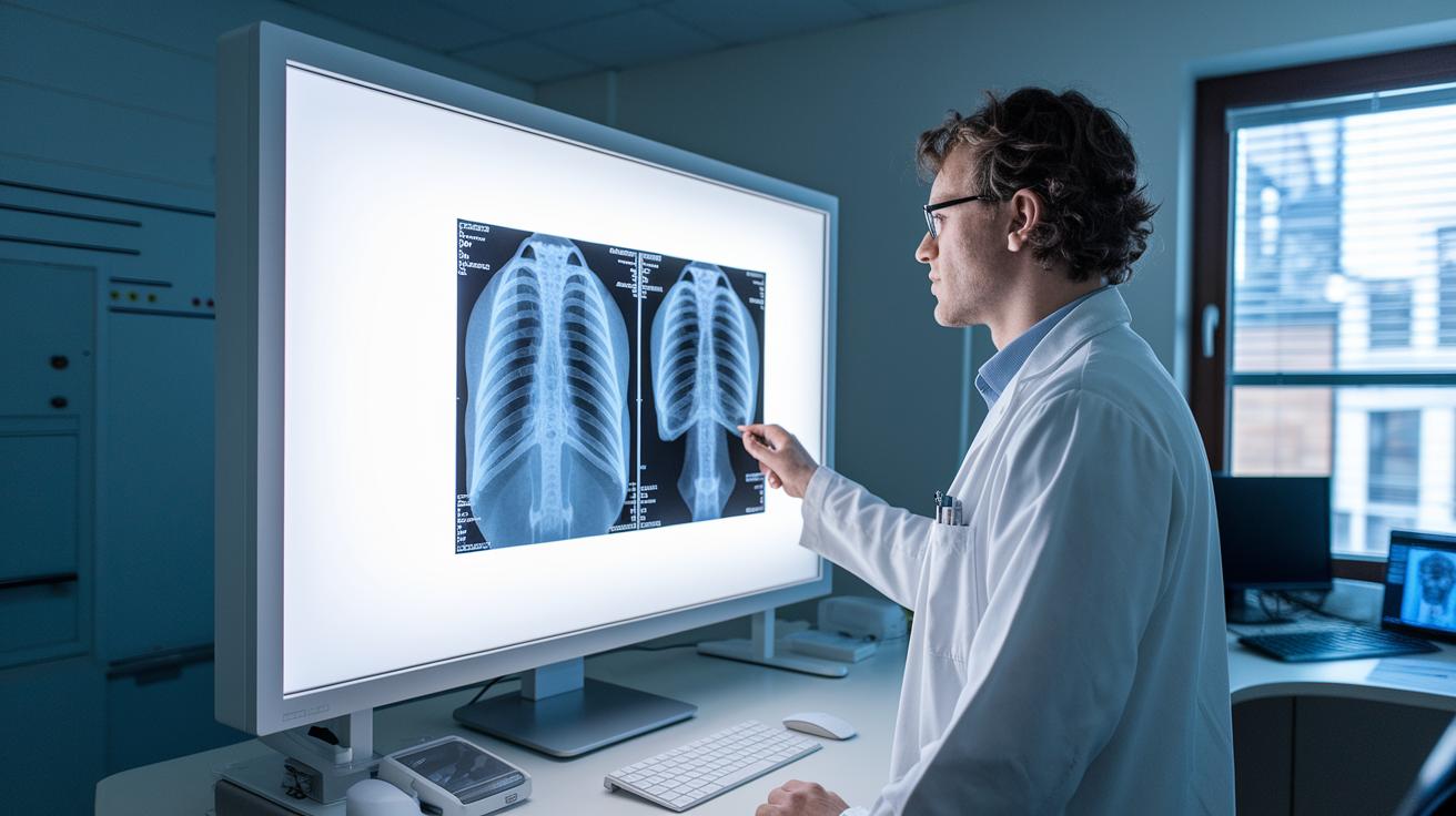

Doctors now can see inside your body faster thanks to digital imaging. This new method gives clear, sharp pictures instantly, which helps doctors make quick decisions.
Traditional film methods have been trusted for years, but they take more time to develop. That extra time can slow down a diagnosis and delay treatment.
Digital imaging speeds things up by capturing images in seconds and storing them electronically. This fast, clear view can lead to more timely care and may improve outcomes for everyone.
Digital Medical Imaging vs Conventional Imaging: Clear Benefits
Digital imaging uses modern detectors that change X-ray energy into electrical signals. The result is an image in seconds, which helps doctors quickly check for injuries and internal issues. This speedy process improves care by giving fast and precise results.
Traditional imaging uses film and chemicals to create pictures. Although film has provided detailed views for many years, it takes longer to produce an image and the quality can vary. Because of these limits, many places are switching to digital methods.
| Aspect | Digital Imaging | Conventional Imaging | Implications |
|---|---|---|---|
| Image Capture | Uses digital detectors to convert X-ray energy into signals | Depends on film and chemical processing | Faster image capture speeds up care |
| Processing Time | Instant results and review | Takes minutes or longer | Quick processing helps in urgent cases |
| Storage | Stored electronically for easy access | Requires physical space and films can degrade | Simplifies record keeping and retrieval |
| Access | Accessed via systems on computers and mobile devices | Relies on physical archives and manual searching | Better access supports remote consultations |
Digital imaging also boosts the clarity of images, showing better contrast and detail. It cuts down waiting times and storage issues. Fast reporting and easy integration with electronic records add extra benefits. These clear advantages make digital imaging a top choice for fast and efficient patient care.
Technical Foundations: Digital Detectors versus Film-Based Radiography
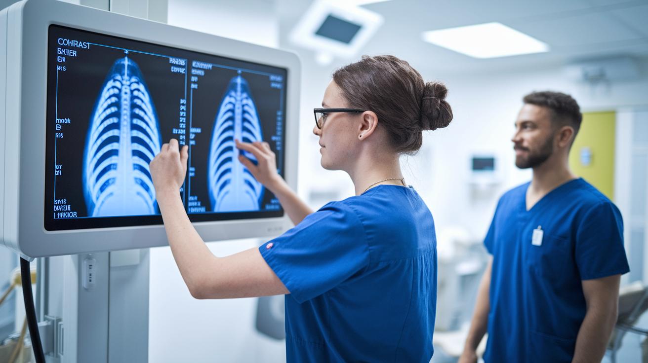
Digital detectors turn X-ray energy straight into electrical signals and create images instantly. This quick process lets technicians adjust settings like contrast and brightness on the spot. It also reduces the need for extra scans and speeds up patient care. For example, digital systems can detect small tissue differences that film might miss without additional exposures.
Film-based radiography, in contrast, captures images on film that must be chemically developed. Although high-quality films can show fine details, they offer a more limited brightness range than digital images. The chemical development process can be sensitive to film quality and timing, which sometimes leads to variations in image consistency and can delay treatment decisions.
Digital systems go beyond traditional X-rays by integrating with other tools such as CT and MRI scans. These methods benefit from digital processing that supports 3D reconstructions and real-time imaging similar to ultrasound, without needing film. Modern image processing lets radiologists fine-tune pictures after capture, highlighting key clinical details. This streamlined digital approach improves diagnostic accuracy and helps clinicians provide faster, patient-centered care.
Image Quality and Diagnostic Accuracy in Digital vs Conventional Imaging
Digital radiography now delivers sharper, clearer images than old film methods. Modern digital detectors capture pictures with a wide range of brightness and better contrast. This means fewer repeat scans and helps doctors spot even tiny problems.
Real-time ultrasound and clearer CT or MRI scans are additional perks of digital systems. These tools provide steady resolution and reduce the unpredictability that sometimes happens with film-based images. In simple terms, digital imaging offers more reliable data for spotting injuries and diseases.
| Feature | Benefit |
|---|---|
| Resolution | Shows finer details in tissues |
| Contrast | Makes image differences clearer |
| Sensitivity | Detects issues well even at lower doses |
| Dynamic Range | Captures subtle brightness changes |
| 3D Reconstruction | Provides detailed cross-sectional views |
Digital methods boost diagnostic accuracy by pairing quick image capture with on-the-spot adjustments. Radiologists can tweak brightness and contrast right after the scan, meaning fewer retakes. The chance to build 3D views from CT or MRI scans adds extra information for a complete check-up. This smooth workflow not only speeds up decision-making in clinics but also leads to clearer and more reliable diagnoses, which ultimately helps guide timely treatments.
Operational Efficiency and Cost Considerations in Imaging Modalities
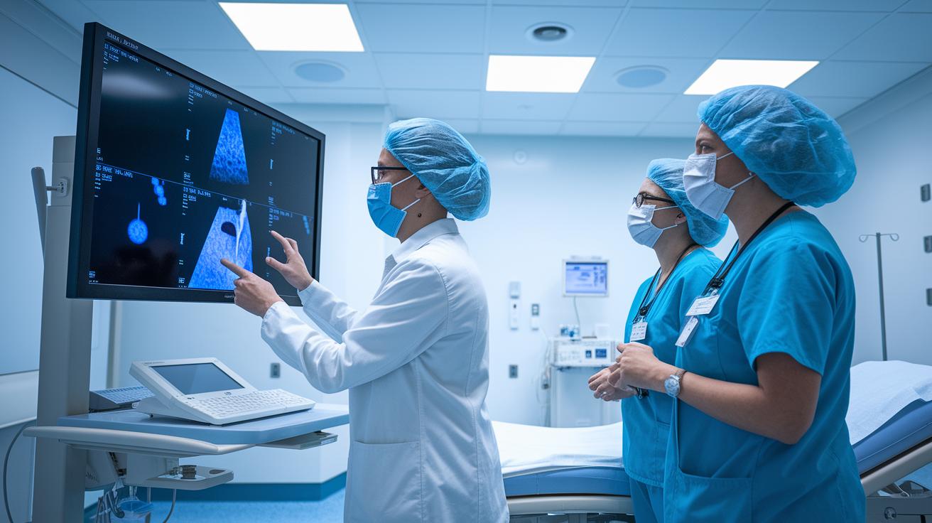
Digital imaging makes work faster and smoother for clinics and patients. Digital detectors capture images in seconds so radiologists can get instant feedback and quickly prepare reports. This speed helps clinical teams decide on treatment faster and see more patients without delay. For instance, during a critical procedure, a digital system delivers an image almost immediately, making timely treatment possible. The seamless integration with electronic records also boosts overall patient care.
Film-based imaging takes longer because it involves several steps before the final image is ready. Traditional methods use chemicals to develop film, which can turn minutes into hours. In urgent situations, these delays are especially problematic. Film also requires extra storage space and extra administrative work to manage physical archives. Moreover, the quality of film images might degrade over time, leading to less reliable service compared to digital systems.
When planning for future investments, digital imaging is more financially sensible despite its higher initial cost. Eliminating recurring expenses for film and chemicals can save money in the long run. Lower material costs and a faster workflow free up resources for other clinical needs. Investing in digital technology not only improves the speed of diagnoses but also reduces extra costs associated with storage and chemical disposal. In the end, digital systems offer a balanced approach that improves patient care while keeping costs under control.
Radiation Exposure and Safety Standards in Digital and Conventional Imaging
Digital radiography uses advanced detectors that capture X-ray signals efficiently. This means a lower dose of radiation is needed to get a clear picture. Technicians can check the image right away, reducing the need for extra shots. This quick feedback helps everyone follow current safety rules and lowers risk for both patients and staff. In addition, digital systems eliminate the need for chemicals used in film processing, which is better for the environment.
Conventional film imaging, by contrast, uses higher radiation levels to produce clear film. The film development process can force extra exposure if retakes are needed, raising risk. Plus, the chemical waste from film processing must be handled with care to protect the environment. In short, digital imaging offers safer, cleaner imaging by lowering both radiation exposure and chemical hazards.
Integration, Artificial Intelligence, and Future Trends in Imaging Technology

Digital picture archives now connect smoothly with electronic health records. Doctors can quickly access scans and images on computers, tablets, or smartphones. This means that a radiologist can check a scan on a tablet during a consultation, making it easier to get the right patient details fast and improving care.
Machine learning and AI are also helping to speed up many routine tasks in radiology. For example, AI tools can automatically measure areas in a scan or highlight regions that look unusual. This can reduce the time doctors spend on manual review and help cut down on errors, leaving more time for solving tougher diagnostic puzzles.
Looking ahead, new research promises even more exciting changes. Advancements in 3D imaging for CT and MRI scans are set to give even clearer views, and breakthroughs in ultrasound may offer detailed real-time images. As AI continues to improve, doctors might soon benefit from better predictive tools and decision support, making diagnoses quicker and turning complicated cases into more manageable challenges.
Final Words
In the action, we examined how digital medical imaging vs conventional imaging differs in key areas. The post broke down modern detector technology, image clarity, workflow efficiency, and safety measures. It also compared integration with AI and emerging trends that are shaping diagnostics. Each method offers distinct pros and cons that can impact care decisions. Advances in digital techniques pave the way for faster, safer patient evaluations, giving room for improved confidence in daily diagnostic practices.
FAQ
Q: What is the difference between conventional radiography and digital radiography?
A: The difference is that conventional radiography uses film and chemical development while digital radiography employs electronic detectors for quick image capture and simpler storage.
Q: What are the 4 types of medical imaging?
A: The four types typically include X‑ray, ultrasound, computed tomography (CT), and magnetic resonance imaging (MRI), each using distinct technologies to create body images.
Q: What imaging examples are considered diagnostic imaging?
A: Diagnostic imaging examples include X‑rays, CT scans, MRIs, ultrasound, and nuclear medicine tests, which all help clinicians assess internal body structures and conditions.
Q: What are the pros and cons of digital imaging equipment?
A: Digital imaging equipment offers benefits like faster image review, lower radiation doses, and easier data storage, while drawbacks include higher initial costs and reliance on advanced technology.
Q: How does conventional radiography compare to computed radiography?
A: The comparison shows that computed radiography uses digital plates for capturing images with quick processing, whereas conventional radiography relies on film that requires longer chemical processing times.
Q: What are the 5 imaging techniques?
A: The five common imaging techniques are X‑ray, computed tomography (CT), magnetic resonance imaging (MRI), ultrasound, and nuclear medicine, each serving different diagnostic purposes.
Q: What is the safest form of medical imaging?
A: The safest form is usually ultrasound, as it does not use ionizing radiation and is often preferred for examining sensitive groups like children and pregnant people.
Q: Why are lead aprons no longer recommended?
A: The reason is that modern imaging methods reduce the need for extra radiation protection, and updated safety practices now favor more precise measures to manage exposure during exams.
Q: Which advantage does digital imaging have over conventional imaging?
A: Digital imaging provides rapid image access, improved workflow through immediate review, and easier sharing and storage, reducing the likelihood of repeat scans and delays in care.