
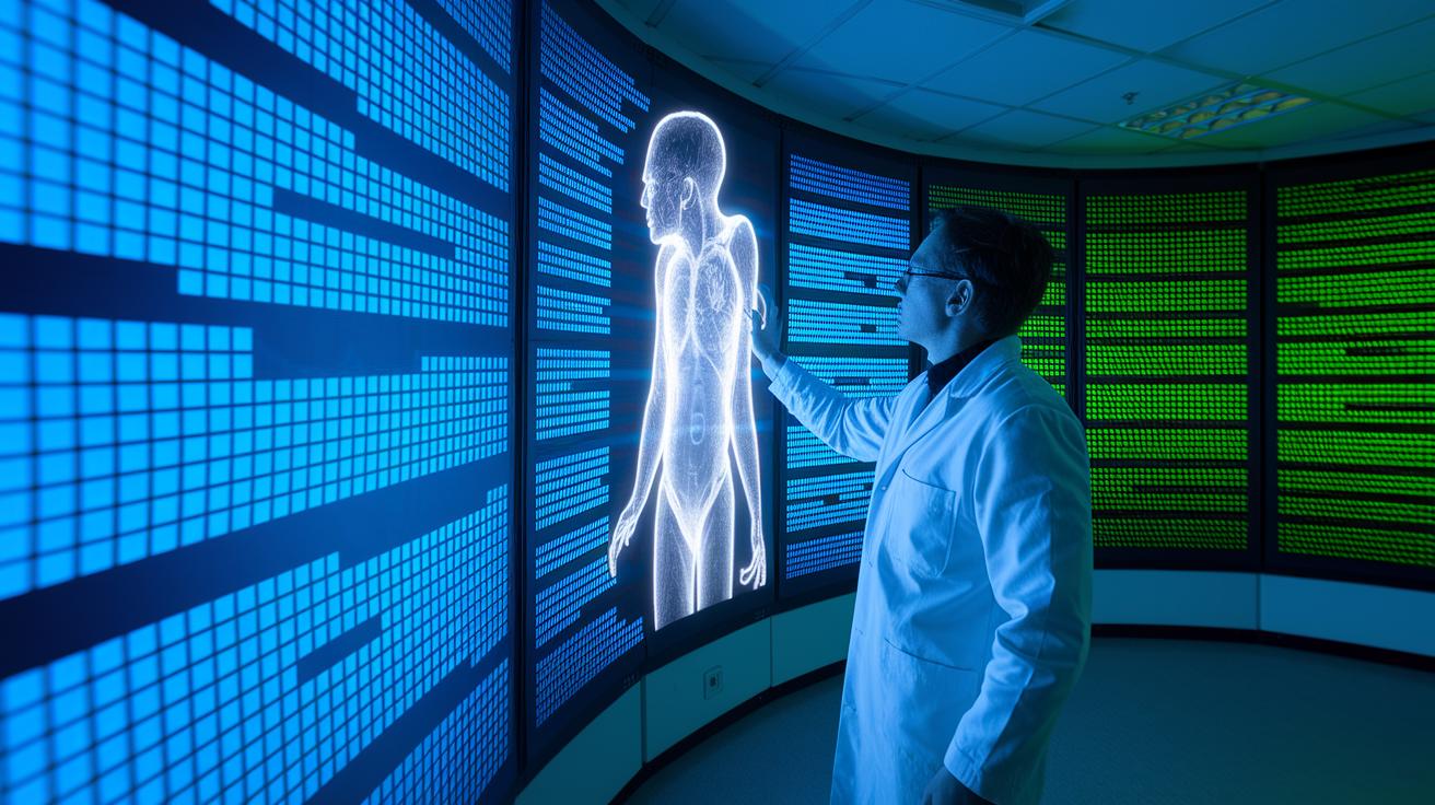

Have you ever wondered how computer programs can uncover hidden details inside your body? Modern medical imaging uses smart software to enhance and break down scans from X-rays, MRIs, CT scans, and PET scans. These programs help doctors see the difference between a tumor and healthy tissue so they can plan treatment with care. By combining artificial intelligence and computer vision, this technology makes sense of complex scans and helps improve patient care.
How Medical Image Analysis Works: Tech meets clarity
Medical imaging uses tools like X-ray, MRI, CT, and PET scans to take pictures of the inside of your body. New software uses advanced computer techniques such as machine learning and deep learning to review these images quickly and accurately.
The process starts with cleaning up the image. Techniques that reduce noise clear random specks, while contrast enhancements make details easier to see. Other methods remove unwanted signals. These steps leave the images clear and ready for further review.
The next step is image segmentation, which breaks up a scan into important parts. For example, computer programs can separate a tumor from healthy tissue, so doctors can plan treatments with precision. After that, the images are aligned using spatial registration. This means images taken at different times or with different machines line up correctly so the same body part is compared accurately.
Once aligned, the images are turned into a three-dimensional model. This transformation from 2D slices into a 3D view allows doctors to look at organs from various angles and plan surgeries. Sometimes, a two-dimensional view is best for focusing on a specific area for diagnosis. Later, computer programs measure and extract important details, which gives doctors vital numbers to help with decisions.
Artificial intelligence and computer vision work together at every step. They review images much faster than manual methods, helping doctors notice small changes and choose the best treatments. This technology ultimately leads to better diagnosis and care for patients.
Medical Image Segmentation Techniques in Image Analysis
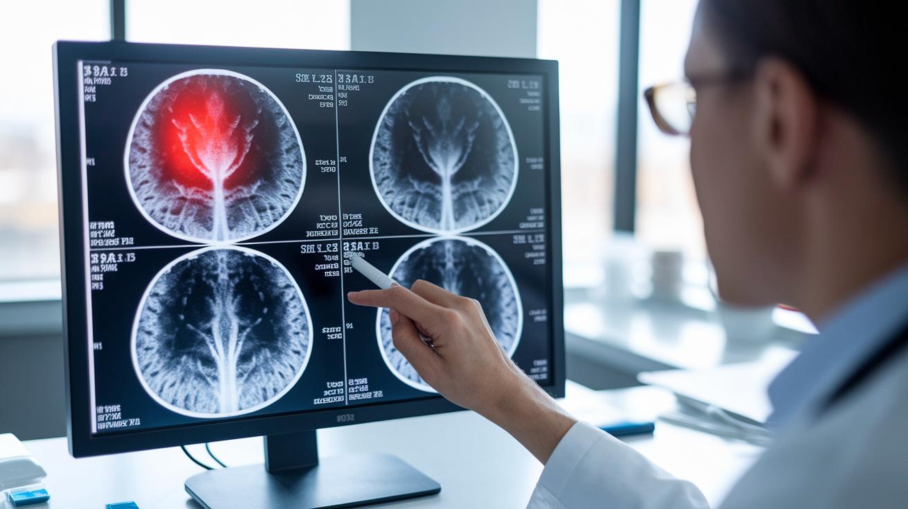
Medical image segmentation is a way to break down scans into clear parts. It lets doctors spot important areas, like separating a tumor from healthy tissue. This step is key when planning treatments, especially in cancer care, because it helps target therapy and set precise doses.
Different methods help create these clear divisions:
- Thresholding methods: This simple approach splits areas based on brightness. For example, it can separate darker tumor parts from lighter tissues.
- Region-growing algorithms: These start at a chosen spot and gradually include nearby, similar pixels, much like water spreading to fill a container.
- Graph-based models: Here, the image is treated like a network of pixels, and the method finds the clear borders between regions.
- Feature-led segmentation: This technique uses details such as texture or shape to identify different tissues, highlighting small differences.
- Deep learning–based segmentation: In this method, a trained neural network learns quickly to tell different regions apart, offering high accuracy.
Each of these methods helps doctors study tissues and plan care, from preparing for surgery to fine-tuning treatment strategies.
Classification and Pattern Recognition in Medical Image Analysis
Image classification algorithms sort through large amounts of scan data and highlight important details for diagnosis. They mark parts of an image as either non-threatening or potentially harmful and point out tiny signs of cancer that might be missed at first glance. For example, when a CT scan shows a small nodule, these tools help doctors decide if it looks like a tumor.
Pattern recognition models add another layer of safety by spotting unusual features, such as small calcium deposits in mammograms or irregular nodules in lung CTs. These systems compare a new image with patterns seen in healthy tissues. When they notice differences, they alert the doctor, suggesting that the area may need a closer look.
Key steps in this process include:
- Using neural networks to study shapes and textures in images
- Sorting parts of an image as likely to be cancerous or not
- Flagging even small deviations with anomaly detection methods
These techniques speed up the review process and help doctors focus on areas that need more attention. Overall, pattern recognition turns complex medical images into clear, actionable information, paving the way for earlier and more effective treatment decisions.
Feature Extraction and Quantitative Biomarker Computation
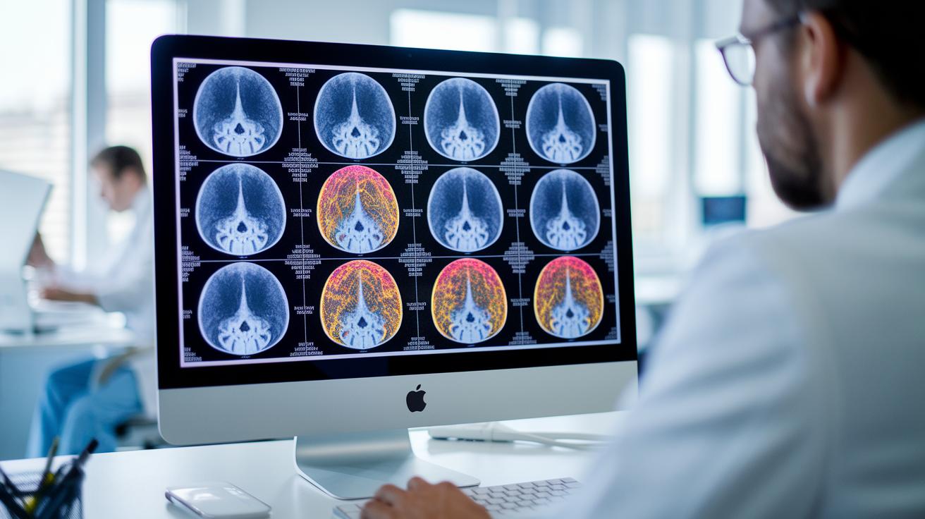
Radiomic workflows analyze images by looking closely at texture, shape, and brightness. These measured traits, called quantitative biomarkers, help doctors connect scan details with a patient’s diagnosis, outlook, and treatment options, especially in cancer and heart care.
There are different methods used to pull features from images:
| Method | What It Does |
|---|---|
| Histogram-based metrics | This approach measures how brightness is spread out. For example, a histogram might show an unusual range of brightness in a suspected lesion. |
| Texture analysis | This technique reviews patterns in pixel brightness. It can reveal, for instance, that a tumor’s texture is rougher than that of normal tissue. |
| Morphological descriptors | These markers describe the shape and size of structures in an image, which may indicate if a mass is likely benign or malignant. |
| Wavelet transforms | This method breaks down an image into different frequency levels to expose small changes in tissue properties. |
By using these biomarkers, clinicians get measurable clues about tissue behavior. This approach connects detailed computer analysis with everyday medical decisions to support personalized care.
Multimodal Data Fusion and Three-Dimensional Reconstruction in Medical Imaging
Combining scans from CT, MRI, PET, and ultrasound helps doctors see both body structures and how they work. This method, called multimodal data fusion, mixes different images to build a fuller picture. By merging these scans, doctors can spot details that one type of image might miss.
Once the images are fused, flat two-dimensional slices are turned into 3D models. These models let clinicians explore organs from all angles. For example, a surgeon might rehearse a procedure on a 3D model before performing the actual surgery, which helps improve planning and safety.
The next step is to analyze the 3D data. This process calculates measurements like organ volumes, tumor shapes, and even how radiotherapy doses spread. These numbers are useful for tracking a disease's progress and checking how well a treatment is working.
| Imaging Modality | Fusion Technique | Clinical Application |
|---|---|---|
| CT & PET | Combined metabolic and anatomical mapping | Tumor staging and treatment guidance |
| MRI & Ultrasound | Overlaying high-contrast soft tissue details | Surgical planning and biopsy targeting |
| CT & MRI | Integrative spatial registration | Pre-operative planning and precision therapy |
These advanced fusion techniques and 3D models are key tools in modern healthcare, helping tailor treatment plans to each patient’s needs.
Challenges and Emerging Trends in Medical Image Analysis
Medical image analysis struggles with issues like keeping patient data private, checking that models work well, and fitting new software into systems already in use. To build trust in these tools, clear ethical rules and guidelines are needed. These rules help stop biased results and protect patient information while new imaging technology continues to improve.
Many health systems use different software platforms, which makes it hard to connect new diagnostic tools smoothly. Cloud solutions and integrated platforms can help manage the storage and computer power needed for large, high-quality imaging data. This makes it easier to share and look at information between hospitals and research centers.
New research in unsupervised learning and deep neural networks is promising better accuracy in image analysis. These advances could help doctors find diseases sooner and more accurately, making important diagnostic tools available to more people. At the same time, the global market for AI-driven imaging solutions is growing fast, AI tools in imaging are expected to grow about 26% annually, while overall image-analysis software is growing by about 8.1%. As these trends continue, both developers and clinicians work to balance cutting-edge innovation with patient safety and data security.
Final Words
In the action, this article showed how medical image analysis works by breaking down each step, from noise reduction and contrast enhancement to segmentation and three-dimensional reconstruction. It explained how AI and computer vision improve detection through pattern recognition and quantitative biomarkers. We also touched on challenges like data privacy and emerging trends in integrating multimodal imaging. The discussion offers a clear look at current techniques that can help shape future patient care and support informed health decisions with optimism about innovations ahead.
FAQ
What does deep learning in Medical image analysis PDF cover?
The deep learning in Medical image analysis PDF covers how neural networks and algorithms enhance tasks like segmentation, classification, and quantification in imaging, improving speed and diagnostic accuracy.
What do Artificial Intelligence in medical imaging PDF documents detail?
The Artificial Intelligence in medical imaging PDF documents detail how AI techniques support image preprocessing, segmentation, anomaly detection, and quantitative analysis, providing insights into improved diagnostic approaches.
How can one access free AI medical image analysis resources?
Free AI medical image analysis resources offer open-access papers, study guides, and sample datasets, enabling students and professionals to explore AI applications in imaging without cost.
What does an AI in medical imaging research paper typically discuss?
An AI in medical imaging research paper typically discusses machine learning methods, feature extraction processes, and comparative analyses with traditional techniques to enhance diagnostic imaging.
How is the Medical image analysis Impact Factor determined?
The Medical image analysis Impact Factor is determined by tracking citations of published articles, reflecting the journal’s influence and the quality of its contributions to imaging research.
What will I learn in a Medical image analysis course?
A Medical image analysis course teaches imaging modalities, preprocessing techniques like noise reduction and contrast enhancement, and AI-driven segmentation and classification methods for clinical use.
What is the focus of AI in medical imaging and diagnostics?
AI in medical imaging and diagnostics focuses on automating image processing and pattern recognition to improve diagnostic precision and efficiency in clinical settings.
What are the benefits of deep learning for medical image analysis?
Deep learning for medical image analysis offers benefits such as enhanced segmentation accuracy, improved anomaly detection, and faster processing, leading to more reliable diagnostic outcomes.