


Have you ever felt confused by your radiology report? You’re not alone. Many patients struggle with the medical terms and numbers that fill these documents.
In reality, a radiology report is split into five clear parts. First, it explains what the images show. Next, it gives detailed results in an easy-to-read format. Another section compares your findings to normal ranges, and a fourth part looks at any changes from previous tests. Finally, the report offers guidance on what to do next.
This guide breaks down each section in simple terms. With this approach, you can feel more confident when reading your exam details and discussing them with your doctor.
Quick Roadmap to a Radiology Report’s Five Key Sections
Radiology reports use a simple, organized format that helps doctors understand what the images show. Each report usually includes five sections that give a full picture of the exam.
• Indication
This section explains why the exam was needed. It describes things like the patient’s symptoms, past diagnoses, and overall clinical concern. For example, a report might say, "Patient experiencing persistent abdominal pain with suspected appendicitis." This helps set the stage for what the doctor is looking for.
• Technique
Here, the report details how the images were taken. It lists the type of test used, whether it's an X-ray, CT, ultrasound, or MRI, and notes details like slice thickness or if a contrast dye was used. An example could be, "CT scan performed with 5 mm slices and contrast enhancement." This information helps doctors understand the quality and limitations of the images.
• Comparison
In this part, the radiologist mentions any previous imaging studies and their dates. Seeing past images side-by-side helps detect changes in a patient’s condition over time. For instance, a report might include, "Compared with the MRI from 02/15/2022, no significant differences are noted." This makes it easier for doctors to track progress or changes.
• Findings
The Findings section is a detailed description of what was seen on the images. It points out normal structures and any unusual areas, such as a "small lesion in the upper lung field." These details form the basis for further diagnosis and treatment.
• Impression
The final section wraps everything up with a short summary of the radiologist’s observations and initial thoughts on the next steps. An example might be, "Observed changes are consistent with early signs of pneumonia." This summary gives the doctor a clear takeaway and guides further discussion or action.
Each section builds on the one before it, creating a step-by-step guide that makes the report clear and easy to follow.
Reading the Indication and Clinical Information in a Radiology Report

The Indication section explains why the study was ordered. It lists the patient's symptoms, past diagnoses, and any referral notes. For example, you might see a note that says, "Patient experiencing sharp chest pain with a history of cardiac issues," which sets the focus for the imaging test.
For a closer look, pay attention to subtle language cues that can give extra insight into the patient's condition. A phrase like "ongoing mild discomfort" might point to a long-term issue rather than an emergency. Also, check if the clinical history mentions minor or occasional symptoms that could affect how the radiologist reads the images.
Review the report header for important details such as the patient's name, the exam date, and the imaging method used (like CT, MRI, or ultrasound). These details provide context that helps you better understand both the Findings and Impression sections.
Key details to note:
- Patient symptoms and previous diagnoses
- Referring physician’s notes
- Examination date and imaging method
Additional tips for in-depth interpretation:
- Look for descriptive terms; for instance, "recurrent headaches over 6 months" suggests a pattern that may need comparison with older reports.
- Watch for minor changes in clinical language that could indicate a shift in the patient's condition.
These insights help turn a basic overview of the test’s purpose into a more detailed understanding of the patient's medical history.
Understanding Technique and Comparison Sections in Your Radiology Report
The Technique section explains how your images were taken. It lists the type of scan, how the images were captured (like slice thickness), and whether a contrast dye was used. These technical details affect what doctors see in the images. For example, a CT scan with 3 mm slices can show smaller spots better than one with 5 mm slices. A case study found that a CT with thinner slices picked up a small lung nodule that thicker slices missed. Every setting, from when the contrast is used to how thick the slices are, can change how clear the image is and how easily tiny problems are seen.
The Comparison section looks at any earlier images you had, noting the type of scan and the date when it was done. When different types of scans are used, comparing them can be tricky. For instance, comparing an ultrasound to an MRI might hide details about how a condition is changing. A report might say, "Compared with a CT from 06/15/2020, the MRI on 05/20/2021 shows some change, but because the scans are different, these findings need careful review." Keeping comparisons as similar as possible helps ensure that progress is measured correctly.
Key details to remember:
- Check the technical settings, as they affect image clarity and how confident doctors are in the diagnosis.
- When possible, compare scans that use similar methods to avoid confusion.
Navigating Findings and Impression Sections in a Radiology Report
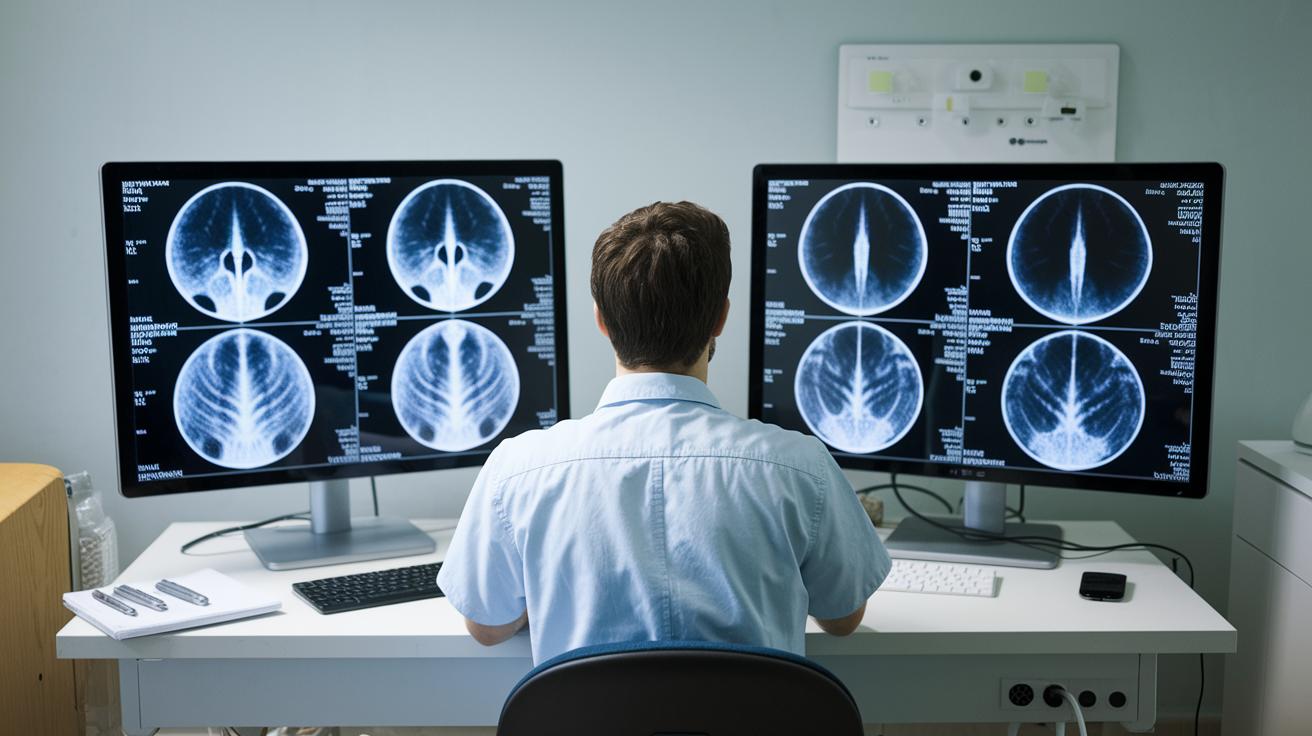
Radiologists split their reports into two main parts. The Findings section lists everything they observed during the scan, from normal structures to potential issues, and uses clear landmarks to show where problems are located. The Impression section then sums up the key points in simple terms, like "Changes are consistent with early inflammation," to highlight what matters most.
For a smooth read, try these steps:
- Start with the Impression to get the main idea.
- Go back to the Findings to see detailed notes and understand how they relate to the summary.
- Watch for clear language such as "increase in size" or "area of concern."
- Note the landmarks mentioned to locate the issue.
For instance, if the Findings mention a "small area of abnormal density in the lung" and the Impression says it is "consistent with early changes," you can easily connect the detailed observation to the radiologist’s main takeaway.
Common Radiology Terms and Abbreviations Explained
Radiology reports can be confusing, but understanding the key words can help you follow along. Here’s a simple glossary to clear things up:
- Hypoechoic: This term is used on ultrasound images when certain areas appear darker. These spots might show fluid or softer tissue.
- Hyperdense: Areas that appear brighter on CT scans. This brightness can suggest calcifications or bleeding.
- Lesion: A general term for any unusual tissue found in the body.
- Nodule: A small, rounded mass that might be harmless or may need further checks.
- Contrast enhancement: When a dye is injected to make images clearer, helping highlight blood vessels or spot abnormal tissue.
- Artifact: A mark or distortion on an image that does not belong to the actual anatomy.
Common abbreviations include:
- R/O: Means "rule out" or exclude a condition.
- w/: Means "with."
- w/o: Means "without."
This clear glossary helps ensure that important details in the Findings and Impression sections are understood correctly.
Final Words
In the action, we broke down the five key sections in a radiology report, from the indication and technique to findings and impression. We also explained common radiology terms and offered a clear snapshot of each part. This guide shows how to read a radiology report (basics) with clarity, helping you understand and use the report when discussing care options. The steps and definitions provided can boost confidence when sharing and reviewing your imaging results with your doctor.
FAQ
Radiology report example
The radiology report example shows a document divided into key sections: indication, technique, comparison, findings, and impression. This layout guides physicians through clinical context and diagnostic conclusions.
Radiology report terminology
The radiology report terminology includes terms like indication, technique, comparison, findings, and impression. These words define parts of the report that convey why the study was done, how it was performed, and what was observed.
Radiology findings vs impression
The radiology findings detail specific observations made during the exam, while the impression provides a brief summary and overall interpretation. This distinction helps clinicians quickly understand the report’s key points.
Sample Radiology Report PDF
The sample radiology report PDF offers a formatted document example. It displays the typical structure, key sections, and language used in comprehensive imaging reports, guiding both professionals and patients.
What does impression mean on a radiology report
The impression on a radiology report means a concise summary of the main findings and preliminary diagnosis. It captures the overall conclusions based on the detailed observations described in the findings.
Radiology report template
A radiology report template outlines essential sections such as indication, technique, comparison, findings, and impression. It provides a structured format for radiologists to ensure clear communication of imaging results.
What is a radiology report
A radiology report is a document created by a radiologist that explains the results of an imaging study. It includes background information, technical details, detailed observations, and a summary of the overall assessment.
Types of radiology reports
Types of radiology reports vary depending on the imaging modality, such as CT, MRI, or ultrasound. Each report follows a structured format but may differ based on institutional practices and specific clinical needs.
What is the 15% rule in radiology?
The 15% rule in radiology refers to a guideline that estimates lesion size changes to assess progression or resolution. This rule aids in evaluating the clinical significance of imaging findings over time.
What is the rule of 10 in radiology?
The rule of 10 in radiology is a guideline used to evaluate lesion characteristics or risk factors. It provides a framework to assist in making decisions regarding further diagnostic evaluation.
How to structure a radiology report?
A radiology report is structured with sections for indication, technique, comparison, findings, and impression. This format organizes clinical reasons, image details, prior study comparisons, observations, and a final summary.
What do findings mean on a radiology report?
The findings on a radiology report mean a detailed description of what was observed during the imaging study. This section lists abnormalities and important observations that inform the overall diagnostic process.