
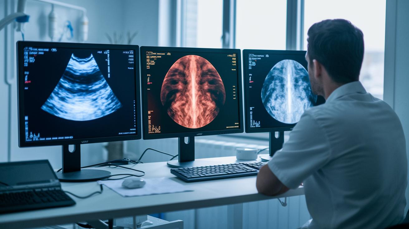

Ever wonder why doctors choose one scan over another? The answer is that each scan has its own strengths.
Ultrasound is safe and shows real-time movement. CT scans give fast, detailed images, though they involve a little radiation. MRI scans take longer but offer clear pictures of soft tissues.
This guide breaks down these methods to help you understand which one might be best in different situations.
Comparative Overview of Ultrasound, CT, and MRI Differences
Ultrasound, CT, and MRI all help doctors view the inside of your body, but each does it in a different way. Ultrasound sends high-frequency sound waves into the body to create live images without using any radiation, making it quick and safe especially for watching moving organs. CT scans, on the other hand, take several X-ray pictures during one breath and combine them into detailed cross-sectional images, even though they use a small amount of radiation. MRI uses strong magnets and radio waves (pulsed radio frequencies) to generate very detailed images of soft tissues, though the scan can take between 15 and 60 minutes to complete.
For urgent cases that need clear, detailed images, CT is usually the option of choice. Ultrasound is great when doctors need to see real-time movement and want to avoid radiation, while MRI shines in offering high-detail images of soft tissues despite its longer scan time. Choosing the best method depends on the patient's situation and the kind of information the doctor needs.
| Modality | How It Works | Scan Time | Best Image Type | Radiation |
|---|---|---|---|---|
| Ultrasound | High-frequency sound waves reflect off tissues | Real-time | Soft tissues | None |
| CT | Multiple low-dose X-rays | Seconds | High-resolution, cross-sectional | Ionizing radiation |
| MRI | Strong magnets and radio frequency pulses | 15–60 minutes | Excellent for soft tissues | None |
Ultrasound Imaging Principles and Clinical Applications

Ultrasound imaging uses high-frequency sound waves that bounce off the body's tissues to create live pictures. This real-time view helps doctors quickly see what’s happening inside.
The image quality depends on the probe's frequency and the skill of the person running the machine. Because ultrasound does not use ionizing radiation, it is safe for pregnant patients and for exams that need to be repeated. Its portability also makes it a great choice for emergency rooms and other point-of-care settings.
Ultrasound exams are used in several key areas:
| Application | Description |
|---|---|
| Fetal Monitoring | Tracks a baby’s heartbeat in real time to spot early signs of distress |
| Cardiology | Shows live heart movements to help identify irregular contractions |
| Trauma Evaluations | Guides emergency assessments by quickly checking for internal injuries |
| Musculoskeletal Imaging | Provides views of muscles and joints to diagnose injuries |
| Abdominal Inspections | Examines organs like the liver and kidneys for abnormalities |
This simple, safe, and flexible method makes ultrasound a valuable tool in many medical settings.
Computed Tomography Fundamentals and Emergency Role
CT scans take several X-ray images while you hold your breath to create a detailed cross-sectional picture of your body. They use smart computer programs that quickly piece the images together. This speed and consistency mean that every scan gives doctors reliable details, which is especially important when time is short.
CT scans are the top choice for looking at injuries from trauma. They help doctors quickly see fractures or internal bleeding. Even though CT scans expose you to a small amount of radiation, the clear images they provide often make this risk worth taking. Hospitals use these scans to diagnose urgent injuries and decide on the best treatment.
Trauma Versus Routine CT Protocols
In emergencies, CT scans are done quickly with faster contrast timing and little patient prep. For routine checks, scans are set up for more detailed imaging, using standard contrast and giving the patient more comfort measures.
Magnetic Resonance Imaging Mechanisms and Soft Tissue Detail
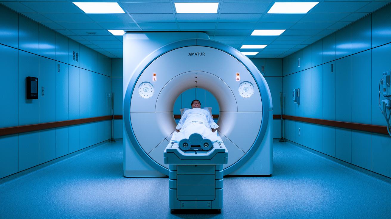
MRI is a scanning tool that uses a strong magnet to line up tiny hydrogen particles in your body, then sends in radio pulses to disturb this arrangement. As the particles return to their normal state, they emit signals that the machine picks up. These signals fade at different rates, which creates a clear picture of various tissues.
This method is particularly useful for examining soft tissues such as the brain, spinal cord, nerves, and tumors. The process provides detailed contrast, which can be the key difference between detecting a tumor or not.
However, there are some challenges. An MRI scan can take anywhere from 15 to 60 minutes, which can be tough for patients who find it hard to stay still. Simple movements or even breathing can blur the images, and metal implants might disrupt the magnetic field. While MRI offers detailed images, it might not work perfectly for every patient or situation.
Safety, Radiation, and Contrast Agent Comparison in Imaging Modalities
When doctors choose an imaging test, they want a safe option that works well for the patient. Each imaging method uses different techniques, and each comes with its own set of benefits and risks.
CT scans create pictures using ionizing radiation. They usually need an iodine-based contrast dye, which can cause allergies or kidney troubles in some people. MRI machines form images with a strong magnet and radio waves, so there is no radiation involved. They use a gadolinium-based dye that is safe for most but can rarely lead to a condition called nephrogenic systemic fibrosis in patients with very poor kidney function. Ultrasound uses high-frequency sound waves to make images and usually does not require any contrast at all, except for a special microbubble agent used in certain blood flow studies.
| Modality | Radiation | Contrast Agent | Main Safety Concern |
|---|---|---|---|
| Ultrasound | None | Rarely used | Minimal risk |
| CT | Ionizing radiation | Iodine-based | Allergic reactions, kidney issues |
| MRI | None | Gadolinium-based | Risk for patients with severe kidney problems |
Doctors decide which imaging test to use based on the medical question and the patient's overall health. For routine checks, the safety of ultrasound is a big plus. However, if faster results or more detailed images are needed, a CT scan or MRI might be the better choice despite their risks.
Cost, Accessibility, and Selection Criteria for Clinical Imaging

Doctors choose between ultrasound, CT, and MRI by weighing practical factors against each patient’s needs. Ultrasound is usually the easiest to get: it costs less, is portable, and doesn’t require special preparation or downtime. CT scans come at a moderate cost and are widely available, with clear safety guidelines for radiation. MRI offers top-notch image detail, but it is pricier, less accessible due to fewer scanners, and often involves longer wait times.
When choosing the best test, clinicians consider these six key points:
- The specific organ system being examined
- How urgent the situation is
- Any patient contraindications
- The cost and potential for reimbursement
- The equipment and resources available at the facility
- The balance of radiation risk versus benefit
This approach helps ensure that the imaging method selected is both effective and safe for each patient.
Final Words
In the action, we explored key points of ultrasound vs CT vs MRI: differences by breaking down physics, scan durations, image strengths, and safety concerns. We also highlighted clinical situations where each imaging option stands out and discussed relevant risks and costs.
This snapshot of comparisons can help guide decisions on selecting the right imaging tool. Keep informed, weigh your options, and remember that your well-being matters every step of the way.
FAQ
Q: MRI vs ultrasound vs CT scan
A: The MRI vs ultrasound vs CT scan comparison shows MRI offers excellent soft tissue detail, ultrasound provides real-time imaging without radiation, and CT scan delivers fast, high-resolution images using ionizing radiation.
Q: Ultrasound vs CT scan vs MRI vs X-ray
A: The ultrasound vs CT scan vs MRI vs X-ray comparison indicates X-rays are best for simple bone imaging, CT scans yield detailed cross-sectional views with radiation, MRI gives superior soft tissue contrast, and ultrasound offers safe, real-time imaging.
Q: MRI vs ultrasound for cancer
A: The MRI vs ultrasound for cancer comparison suggests MRI typically provides higher detail for tumor characterization, while ultrasound can be useful for guiding biopsies and assessing superficial or accessible lesions.
Q: Which is better, MRI or ultrasound?
A: The question of which is better, MRI or ultrasound, depends on the clinical need; MRI offers comprehensive soft tissue contrast, whereas ultrasound is quick, safe, and effective for real-time imaging.
Q: CT scan vs MRI: Which is safer?
A: The CT scan vs MRI safety comparison indicates that MRI avoids ionizing radiation, making it safer for repeated use, while CT scans involve low-dose radiation, though they are valuable in urgent evaluations.
Q: What can a CT scan show that an MRI cannot?
A: The question on what a CT scan shows that an MRI cannot points out CT’s strength in detecting acute bleeding and bone injuries quickly, thanks to its rapid acquisition and use of ionizing radiation.
Q: Stomach ultrasound vs CT scan
A: The stomach ultrasound vs CT scan comparison shows ultrasound offers dynamic, radiation-free imaging ideal for initial screening, while CT scan delivers detailed anatomical views of the stomach and surrounding structures.
Q: Which is better, MRI or CT scan for brain imaging?
A: The MRI vs CT scan for brain imaging question reveals that MRI is preferred for detailed soft tissue analysis, whereas CT scan is often used in emergencies to quickly detect bleeding and fractures.
Q: Which is better, CT scan, MRI, or ultrasound?
A: The question of which modality is better—CT scan, MRI, or ultrasound—depends on clinical factors; CT is fast for emergencies, MRI excels in soft tissue detail, and ultrasound is safe and real-time.
Q: What can an ultrasound show that an MRI cannot?
A: The question on what an ultrasound shows that an MRI cannot highlights ultrasound’s ability to provide real-time imaging, assess blood flow, and guide procedures without using radiation.
Q: Which scan is most accurate?
A: The inquiry about which scan is most accurate depends on the target; MRI is highly accurate for soft tissue detail, CT scans excel in bone evaluation, and ultrasound is best for dynamic assessments in real time.
Q: What does ultrasound show that CT doesn’t?
A: The comparison of what ultrasound shows that CT doesn’t emphasizes ultrasound’s live imaging capability, especially useful in assessing moving structures and blood flow without radiation.