


Ever wonder how doctors can look inside you without surgery? They use medical imaging, a safe and quick way to see what’s happening beneath your skin. Tools like X-rays, ultrasounds, CT scans, and MRIs create clear images of bones, organs, and tissues. These images help doctors spot problems early, from broken bones to infections, so they can decide the best treatment. In short, medical imaging reveals hidden health issues and guides care.
3. what is medical imaging: Clear and Simple
Medical imaging lets doctors look inside your body without surgery. These techniques create pictures of bones, organs, and tissues using tools like ultrasound, X-rays, CT scans, and MRI. For instance, an X-ray can quickly reveal a broken bone, while an MRI shows detailed images of soft tissues. Think of it like taking a snapshot of what’s happening inside using light or sound waves.
These safe tests help doctors decide what might be wrong when you have pain or unusual symptoms. They choose the right method for each situation. For example, an ultrasound is often used during pregnancy to safely check on the baby. CT scans offer many detailed images for suspected internal injuries, and MRI gives a closer look at soft tissues when needed. This careful matching of tests to symptoms means that doctors can find the right answers faster.
By using medical imaging, doctors get a clear view that regular exams can’t provide. It helps them spot problems early, from broken bones and infections to tumors and other issues, so patients can get the treatment they need quickly. Imagine looking at a city map to find a missing street; medical imaging gives doctors the detailed guide they need to pinpoint and treat health problems effectively.
Evolution and Milestones in Medical Imaging
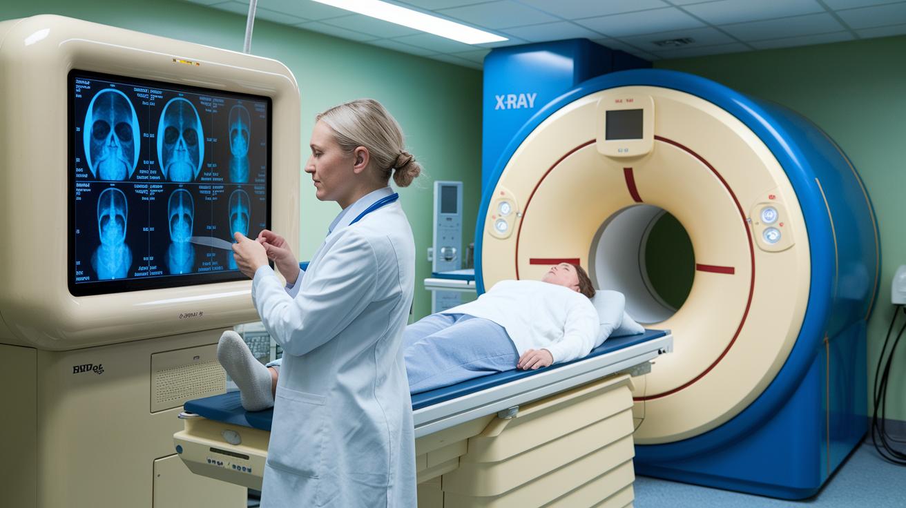
X-rays changed medicine when Wilhelm Röntgen discovered them in 1895. They let doctors see inside the body quickly and clearly, making diagnosis much simpler.
In the 1970s, computed tomography (CT) made a big difference. A CT scan spins an X-ray beam around the body and takes many pictures. These images are compiled into detailed slices that help doctors locate problems fast.
The 1980s introduced magnetic resonance imaging (MRI), which uses magnets and radio waves to show soft tissues without using radiation. Not long after, ultrasound provided real-time images with sound waves, a tool especially handy for monitoring pregnancies. Fluoroscopy also emerged as a method to view live movement in the body.
Later improvements, including positron emission tomography (PET) for tracking how the body uses energy, and advanced 3D imaging techniques, have all added to the precision of these tests. Each step in this journey has made it easier for doctors to accurately identify issues and improve patient care.
Key Modalities in Medical Imaging
-
X-rays: X-rays use high-energy waves that pass through the body. Bones soak up more of these waves, so they appear brighter on the film. This method is quick and affordable, which is why doctors often use it to screen for fractures or check for pneumonia. However, since it produces a flat image, some 3-dimensional details might be missed.
-
Ultrasound: Ultrasound sends sound waves from a device into the body. The waves bounce off tissues and return as echoes that form a real-time image. It is safe and non-invasive, which makes it a common choice for monitoring a baby during pregnancy. Yet, ultrasound does not work well with bones or deep tissues, so it is best for imaging areas such as the abdomen or pelvis.
-
CT Scans: CT scans take multiple X-ray images as the machine rotates around the body. These images are then rebuilt into detailed 3-dimensional views. CT scans can help diagnose conditions such as pulmonary embolism or internal injuries. The clear cross-sectional images are very useful, though the procedure uses a higher dose of radiation than regular X-rays.
-
MRI: MRI uses powerful magnets and radio waves to capture detailed images of soft tissues like the brain, muscles, and organs. Often, a gadolinium-based contrast agent is used to improve clarity. A major advantage is that it does not use ionizing radiation. However, the strong magnetic field means it is not suitable for people with certain metal implants.
-
PET: PET scans use radioactive tracers to map cell activity and metabolism within the body. This technique is especially common in cancer care to detect tumors and assess their stage. While PET provides important information about how cells function, it involves radiation exposure and usually offers lower image detail.
-
Nuclear Medicine Imaging: This method also involves radioactive tracers, but it focuses on showing how well organs are working, such as measuring thyroid function or evaluating heart blood flow. Although it helps assess organ performance, the images lack the finer anatomical details seen in some other techniques and involve radiation exposure.
Under the Hood: How Medical Imaging Systems Work
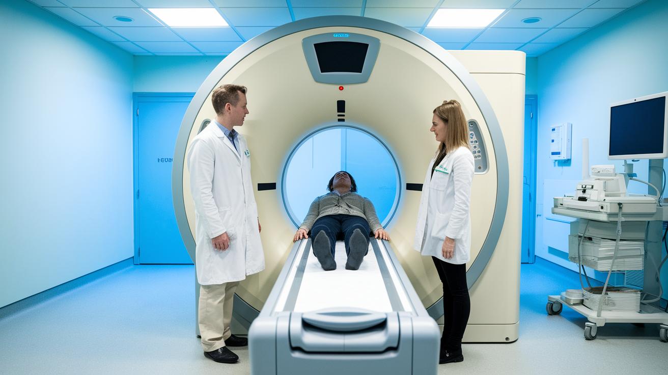
CT and MRI scans start by taking many pictures as the machine moves around the body. Advanced computer programs then stitch these individual slices together, forming a complete cross-section or even a 3-dimensional view. For example, a CT scan spins its X-ray source in a full circle to capture multiple images, which are later combined to give a clearer look at internal structures.
Digital imaging systems play a key role by turning old-style analog signals, like those from X-ray films or ultrasound echoes, into high-resolution digital files. This digital conversion makes it easier and faster for doctors to share and analyze images with their teams.
Then, specialized software boosts image quality by reducing errors and enhancing clarity. This refined image allows clinicians to measure tissue size, density, or identify abnormal areas that could be important for diagnosis.
Contrast agents add another layer of detail. In CT scans, iodine-based agents help highlight blood vessels and tissues, while MRI scans often use gadolinium to shed light on lesions and key boundaries. This boosts a physician’s ability to pinpoint areas of concern with greater precision.
Benefits and Risks of Medical Imaging
Diagnostic tests play a key role in patient care by helping doctors see what's happening inside the body. Each imaging method has its own clear benefits and some risks that are important to consider.
X-rays are quick and affordable. They create a flat, two-dimensional image to spot fractures or lung issues and use only a small amount of radiation.
CT scans produce detailed, layered images that can reveal problems like pulmonary embolism (a blood clot in the lung). They are very fast but involve a higher dose of radiation.
Ultrasound is a safe option because it uses sound waves instead of ionizing radiation. It works well for monitoring a fetus or checking the abdomen, though it may struggle to image deep tissues or view bones clearly.
MRI scans deliver high-detail images of soft tissues without radiation exposure, making them ideal for studying muscles and organs. However, the process takes longer and may not be suitable for patients with metal implants.
Fluoroscopy provides real-time video imaging, which is useful for guiding procedures. It requires cautious use due to the continuous radiation exposure during the exam.
Each of these tests offers different advantages and trade-offs. For example, a CT scan gives more detailed images than an X-ray but comes with a higher radiation risk. Understanding these differences helps both patients and doctors make better choices for care.
Clinical Applications of Medical Imaging
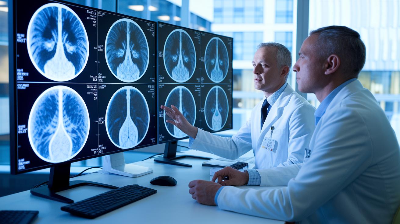
Medical imaging technology is rapidly advancing, making it easier for doctors to diagnose problems accurately. Recent breakthroughs include CT scans that use AI to quickly spot issues like trauma injuries and MRI machines that capture images faster to better assess urgent brain and heart conditions. One case showed an AI system noticing a small lung lesion on a CT scan, which led doctors to take early action.
Traditional imaging methods are also getting upgrades. For instance, ultrasound now offers 3D views to check on baby development more clearly, and PET scans use detailed measurements to stage tumors more accurately. Even fluoroscopy has improved with sharper digital images to help during real-time procedures.
| Modality | Updated Clinical Application |
|---|---|
| X-ray | Detects bone fractures and pneumonia with digital tools that highlight subtle signs |
| Ultrasound | Tracks fetal growth and examines the abdomen using advanced 3D imaging |
| CT Scan | Assesses abdominal pain and trauma with fast, AI-supported diagnostics |
| MRI | Finds soft-tissue issues and evaluates brain and heart conditions with quicker imaging |
| PET | Assists in tumor detection and staging with improved quantitative analysis |
| Fluoroscopy | Supports real-time interventions with clearer digital visuals |
Safety and Patient Care in Imaging Procedures
Getting ready for an imaging test is important for your safety and comfort. Your care team will guide you on what to do before the test, such as fasting or drinking extra fluids for some scans. For an MRI, you will need to remove metal items like jewelry because even small bits of metal can create problems with the strong magnetic field. They will also check if you have any implants or devices that might react to the magnet.
Hospitals and clinics follow strict guidelines to keep radiation doses as low as possible. For example, the ALARA approach (As Low As Reasonably Achievable) is used during CT and fluoroscopy procedures to reduce exposure. Other imaging techniques, like ultrasound and MRI, do not use radiation, though centers still focus on reducing noise and keeping the room comfortable.
Before each test, technicians explain what will happen so you know what to expect. This clear guidance helps reduce any worries and improves test accuracy. You might be asked to stay very still, and some centers even offer earplugs if the scan is noisy. These careful steps help ensure that imaging tests are both safe and effective in providing accurate diagnoses.
Future and Emerging Trends in Medical Imaging
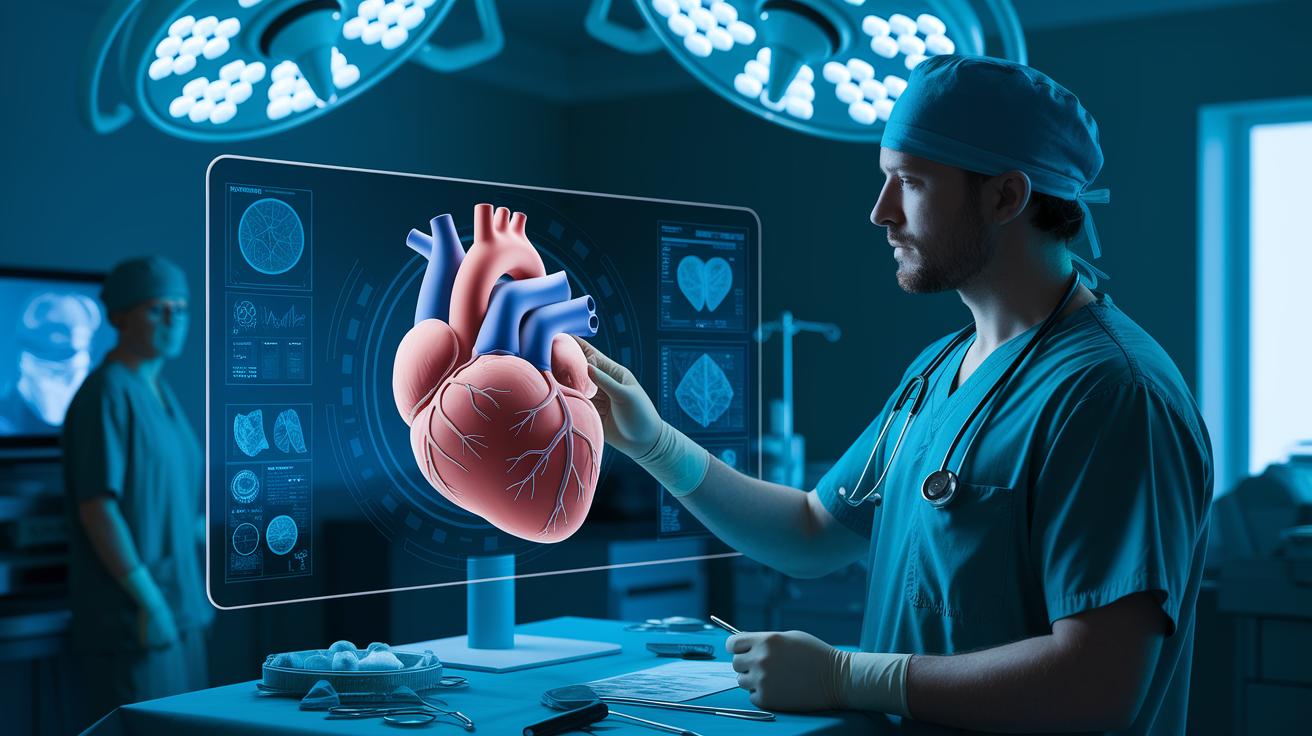
New advances in medical imaging are changing the way doctors plan treatments and guide procedures. With modern 3D imaging, surgeons can now view a detailed digital model of a patient’s heart before entering the operating room. This clear map helps them plan the surgery more carefully.
Real-time imaging is also moving forward. New dynamic scans let doctors watch how organs like the kidney and heart work as they function. This continuous monitoring can catch early changes that might need treatment.
Image-guided procedures have made a big impact as well. Combining live imaging with small incisions allows clinicians to target treatments more accurately while helping patients heal faster. Faster scans with sharper images are making it easier for doctors to diagnose problems correctly.
Linking imaging with digital health tools further enhances patient care. These integrated systems can monitor health changes and adjust treatments to fit each patient’s needs. Overall, these improvements promise better care and a smoother treatment process in clinics everywhere.
Final Words
In the action, the post unpacked what is medical imaging by explaining its basics, how it evolved, and the various tools like X-rays, ultrasound, CT scans, and MRI. It detailed the inner workings of image processing, highlighted both the benefits and risks, and explored real-life clinical applications that support faster, noninvasive diagnoses.
Ongoing trends promise more precise, faster scans and smarter safety protocols. The outlook remains positive, emphasizing continuous improvement and better care for every patient.
FAQ
FAQ
What is medical imaging used for?
Medical imaging is used for capturing noninvasive images of internal body structures. It helps doctors diagnose issues like fractures, tumors, and organ problems using tools such as X-rays, CT scans, MRI, and ultrasound.
What is a medical imaging degree?
A medical imaging degree trains students in diagnostic techniques and technology. Graduates learn to operate imaging equipment and interpret visuals, preparing them for roles in hospitals and clinics.
What is a medical imaging doctor?
A medical imaging doctor, commonly known as a radiologist, specializes in analyzing images like X-rays and MRIs to diagnose conditions and guide treatment decisions.
What is medical imaging technology?
Medical imaging technology covers the systems and devices that create visual representations of the body. This includes X-ray machines, CT scanners, MRI units, and ultrasound systems used for diagnostic purposes.
What are the benefits of medical imaging?
Medical imaging offers benefits such as noninvasive diagnostics, rapid results, and precise detection of abnormalities. It plays a key role in guiding treatment decisions while reducing the need for invasive procedures.
What are the five imaging techniques?
The five primary imaging techniques are X-rays, ultrasound, CT scans, MRI, and PET scans. Each method provides specific insights suited to different diagnostic needs.
What does “medical imaging techniques PDF” mean?
A “medical imaging techniques PDF” is a downloadable document that outlines various imaging methods, their principles, applications, and limitations, serving as a useful study or reference guide.
What is meant by medical imaging?
Medical imaging means creating visual representations of the internal body to assist in diagnosing, monitoring, and treating conditions. These methods offer a noninvasive way to view bones, organs, and tissues.
What are the four main types of medical imaging?
The four main types of medical imaging typically include X-rays, ultrasound, CT scans, and MRI. Each has unique strengths suited to visualizing various internal structures and conditions.
Is medical imaging the same as MRI?
Medical imaging is not the same as MRI. MRI is one form of imaging among other types like X-rays, CT scans, and ultrasound used in medical diagnostics.
What do you do in medical imaging?
In medical imaging, professionals operate and maintain imaging equipment, capture quality images, and collaborate with radiologists to interpret results, ensuring accurate diagnosis and effective patient care.
What is the salary in the medical imaging field?
The medical imaging salary varies by role, experience, and location. Professionals such as radiologic technologists often earn competitive rates that reflect their specialized training and diagnostic expertise.